Copyright
©The Author(s) 2023.
World J Clin Cases. Jan 26, 2023; 11(3): 576-597
Published online Jan 26, 2023. doi: 10.12998/wjcc.v11.i3.576
Published online Jan 26, 2023. doi: 10.12998/wjcc.v11.i3.576
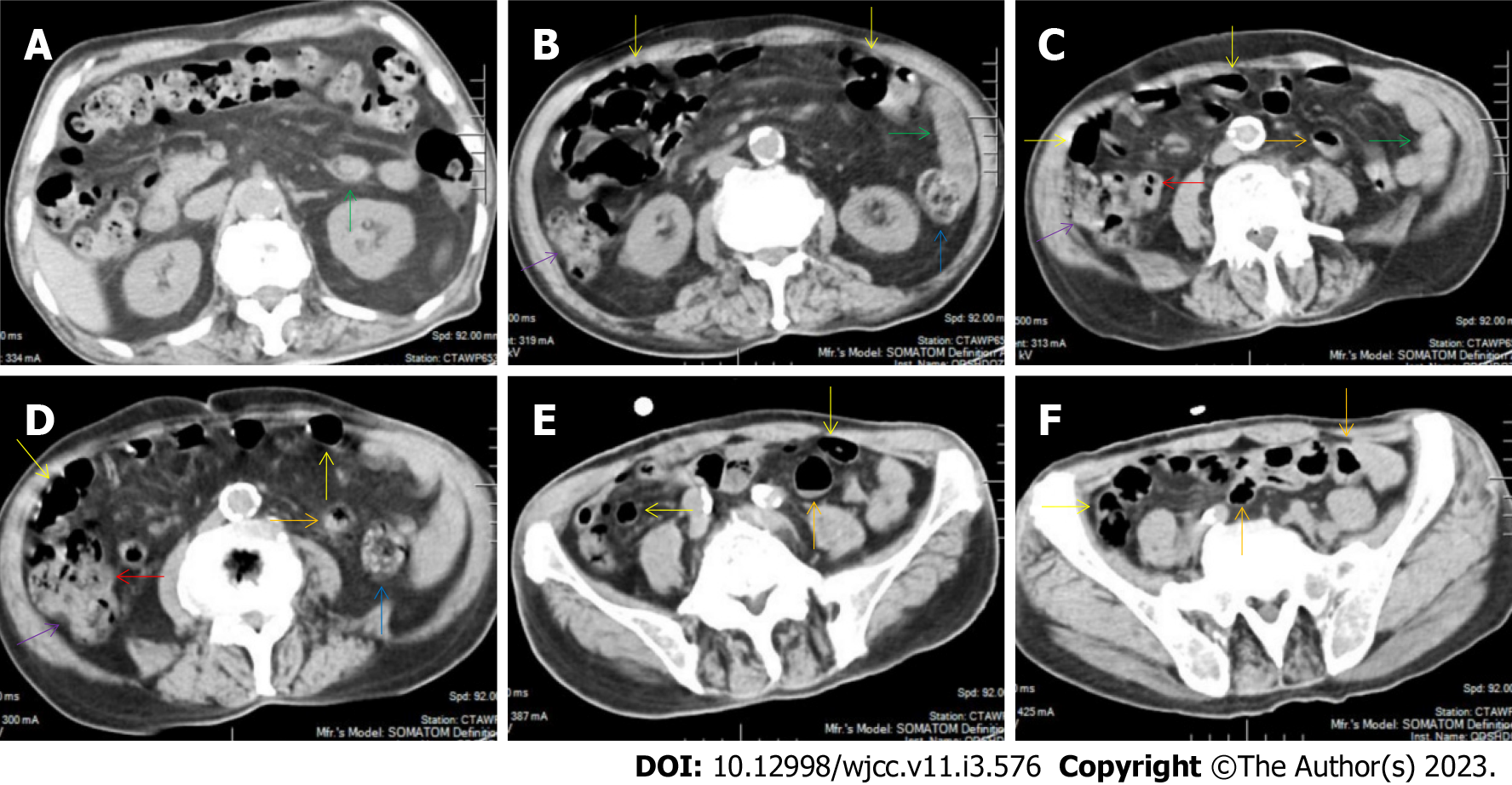
Figure 15 Characteristic images of case 10.
A and B: Characteristic images of the fat deposition. The most noticeable radiological finding was the panabdominal silt-like hypervascular fat deposition, leading to the widening of the bowel loop of the jejunum and the proximal ileum, forming the so-called “creeping fat sign”. The ileum was fibritically thickened and dilated, and the duodenum and the proximal jejunum were liquid-filled (green arrows); C and D: Characteristic images of the ileocecal region. The ileocecal valve and the terminal ileum were significantly thickened, stratified and strictured (red arrows), and proximal to the thickened terminal ileum, the ileal lumen was dilated and gas-filled and the mucosa was hyperdense (yellow arrows). The ascending colonic wall was also thickened (purple arrows). In a short segment of the descending colon (blue arrows), accrescent villi were especially prominent; E and F: Characteristic images of the small intestine. The proximal ileum and the distal jejunum (orange arrows) were thickened and gas-filled.
- Citation: Zhao XC, Xue CJ, Song H, Gao BH, Han FS, Xiao SX. Bowel inflammatory presentations on computed tomography in adult patients with severe aplastic anemia during flared inflammatory episodes. World J Clin Cases 2023; 11(3): 576-597
- URL: https://www.wjgnet.com/2307-8960/full/v11/i3/576.htm
- DOI: https://dx.doi.org/10.12998/wjcc.v11.i3.576


