Copyright
©The Author(s) 2023.
World J Clin Cases. Jan 26, 2023; 11(3): 576-597
Published online Jan 26, 2023. doi: 10.12998/wjcc.v11.i3.576
Published online Jan 26, 2023. doi: 10.12998/wjcc.v11.i3.576
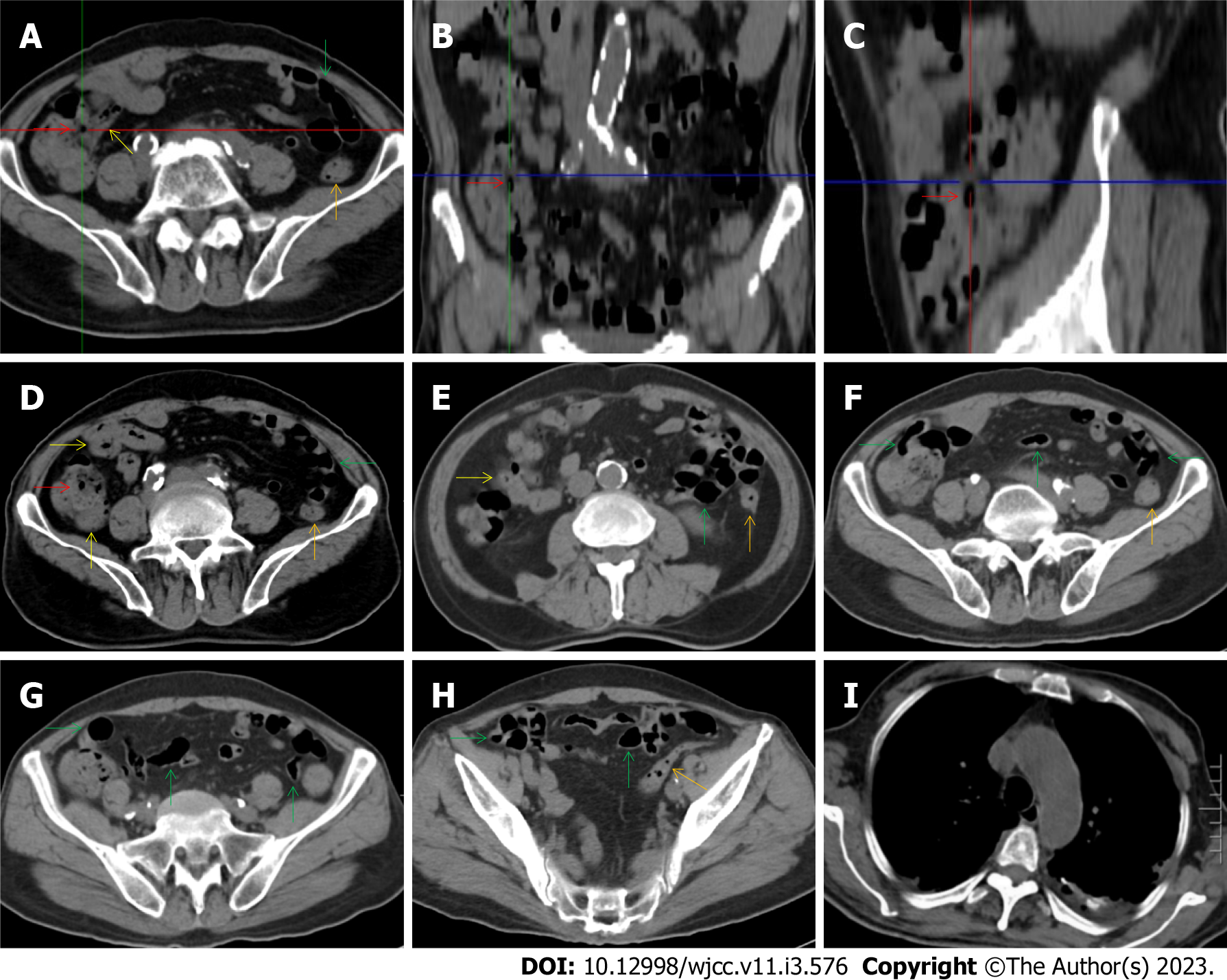
Figure 14 Characteristic images in case 8.
A-C: Characteristic images of the ileocecal region and the small intestine. The ileocecal valve (red arrows) was thickened, strictured and stratified. The distal ileum (yellow arrows) were significantly thickened with intramural gas and wrapped by prominent mesenteric fat stranding, which suggested the presence of aggravated inflammatory damage in the distal ileum and ileocecal region; D-H: Characteristic images of the small intestine. In the ileocecal region, the ileum adhered to the cecum, and the cecum was thickened and stratified. In other colonic segments (orange arrows), the bowel wall was also thickened and stratified, and the lumen was dilated in some segments and collapsed in other segments. From the jejunum to the proximal ileum (green arrows), the wall was fibrotic and thickened and the lumen was gas-filled. Several segments of adhesive bowel loop were present in the small bowel. Noticeably, panabdominal silt-like hypervascular fat deposition wrapped the adhesive and widened small bowel loop (creeping fat sign), which suggested the presence of chronic transmural inflammatory damage and a diagnosis of Crohn’s disease; I: Characteristic image of the chest computed tomography (CT) scan. Chest CT showed exudative lesions in the left pleura in the context of calcified lesions, indicating the reactivation of an old tuberculosis infection.
- Citation: Zhao XC, Xue CJ, Song H, Gao BH, Han FS, Xiao SX. Bowel inflammatory presentations on computed tomography in adult patients with severe aplastic anemia during flared inflammatory episodes. World J Clin Cases 2023; 11(3): 576-597
- URL: https://www.wjgnet.com/2307-8960/full/v11/i3/576.htm
- DOI: https://dx.doi.org/10.12998/wjcc.v11.i3.576


