Copyright
©The Author(s) 2023.
World J Clin Cases. Jan 26, 2023; 11(3): 576-597
Published online Jan 26, 2023. doi: 10.12998/wjcc.v11.i3.576
Published online Jan 26, 2023. doi: 10.12998/wjcc.v11.i3.576
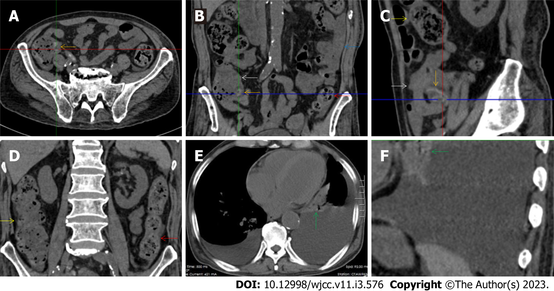
Figure 12 Characteristic images in case 16.
A-C: Characteristic images of the ileocecal region and the small intestine. The ileocecal valve was fibrotic and thickened with edematous fat deposition, forming the so-called “fat holo sign” (orange arrows), and the distal ileum was adhesive with mesenteric fat stranding and peritoneal thickening (white arrow), forming the so-called “abdominal cocoon”. Intestinal adhesion with bowel wall thickening, peritoneal involvement and mesenteric fat stranding was also found in the jejunum (blue arrow). Heterogeneity in the bowel wall texture was particularly prominent in the adhesive bowel segments; D: Characteristic image of the large intestine. The ascending colon was rugged and the colonic wall was thickened and stratified, with adjacent omental thickening (yellow arrows). The descending colonic wall were significantly thickened and stratified with mucosal hyperdensity and paracolonic fat stranding (red arrows); E and F: Characteristic images of the chest computed tomography (CT) scan. Chest CT revealed that the pleural effusion was predominantly in the left cavity and hypertrophic lesions involved both the left pleura and the pericardium (green arrows).
- Citation: Zhao XC, Xue CJ, Song H, Gao BH, Han FS, Xiao SX. Bowel inflammatory presentations on computed tomography in adult patients with severe aplastic anemia during flared inflammatory episodes. World J Clin Cases 2023; 11(3): 576-597
- URL: https://www.wjgnet.com/2307-8960/full/v11/i3/576.htm
- DOI: https://dx.doi.org/10.12998/wjcc.v11.i3.576


