Copyright
©The Author(s) 2023.
World J Clin Cases. Jan 26, 2023; 11(3): 576-597
Published online Jan 26, 2023. doi: 10.12998/wjcc.v11.i3.576
Published online Jan 26, 2023. doi: 10.12998/wjcc.v11.i3.576
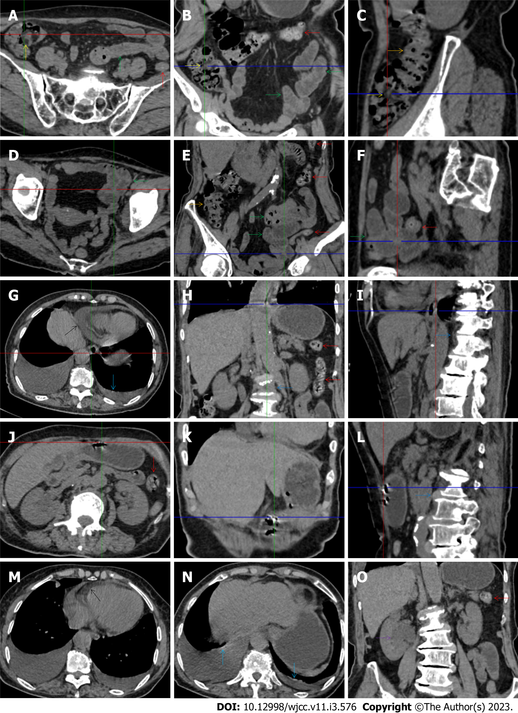
Figure 11 Characteristic images in case 17.
A-C: Characteristic images of the ileocecal region and the ascending colon. Circumferentially distributed fat deposition wrapped the ileocecal valve (yellow arrows), forming the so-called “fat holo sign”. The terminal ileum was thickened, and the strictured ileocecal valve and terminal ileum led to the small intestines being liquid-filled. Heterogeneity and hypertrophic lesions of the small intestines were easily recognized. The ascending colon was rugged with paracolonic fat stranding and peritoneal thickening, and the colonic wall was thickened and stratified with edematous submucosal tissue and intramural gas (orange arrows). From the transverse to the sigmoid colon (red arrows), the colonic wall was fibrotically thickened, with multiple inflamed diverticula in this colonic segment; D-F: Characteristic images of an adhered jejunal segment. A long segment of the adhered jejunal loop was present in the left iliac fossa and pelvic cavity (green arrows). In the adhesive jejunal loop, the lumen was gas-filled in the proximal segment and liquid-filled in the distal segment. Hypervascular mesenteric fat stranding and fibrotic peritoneal thickening were present in this bowel segment, forming the so-called “abdominal cocoon”. Fibrotic wall thickening was present in the entire jejunal segment and accrescent pili were visualized in the adhesive jejunal loop and the proximal jejunum; G-I: Erosive lesions in extraintestinal organs. Erosive lesions on the background of the calcified lesions were found in the esophagus. Hypertrophic lesions and seromembranous effusion were visualized in the pericardium (black arrows) and the pleura (light blue arrows). Exudative lesions were also present in the vertebral column (blue arrows); J-L: Erosive lesions in the stomach. Erosive lesions on the background of the calcified lesions were also found in the stomach; M-O: Exudative lesions in extraintestinal organ. Hypertrophic lesions and seromembranous effusion were visualized in the pericardium (black arrows) and the pleura (light blue arrows). Exudative lesions were also present in the renal pelvis and urinary tract (a purple arrow). These radiological features led to the diagnosis of reactive tuberculosis infections in the gastrointestinal tract, urinary tract, peritoneum, vertebral column, pleura and pericardium.
- Citation: Zhao XC, Xue CJ, Song H, Gao BH, Han FS, Xiao SX. Bowel inflammatory presentations on computed tomography in adult patients with severe aplastic anemia during flared inflammatory episodes. World J Clin Cases 2023; 11(3): 576-597
- URL: https://www.wjgnet.com/2307-8960/full/v11/i3/576.htm
- DOI: https://dx.doi.org/10.12998/wjcc.v11.i3.576


