Copyright
©The Author(s) 2023.
World J Clin Cases. Jan 26, 2023; 11(3): 576-597
Published online Jan 26, 2023. doi: 10.12998/wjcc.v11.i3.576
Published online Jan 26, 2023. doi: 10.12998/wjcc.v11.i3.576
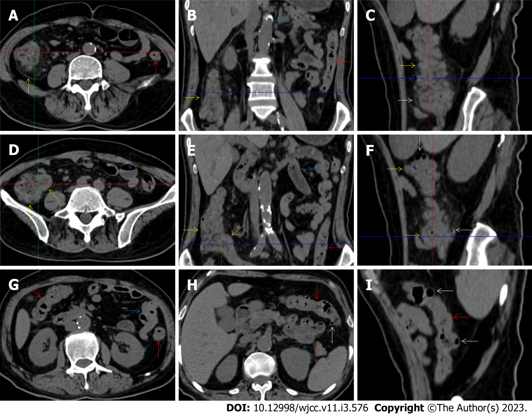
Figure 10 Characteristic images in case 11.
A-C: Characteristic images of the ascending colon. The ascending colon was rugged with fibrotically and irregularly thickened colonic mucosa, circumferentially distributed omental thickening and paracolonic fat stranding (yellow arrows), and other colonic segments were thickened and stratified with water holo sign (red arrows); D-F: Characteristic images of the ileocecal region. The ileocecal valve and the terminal ileal wall were strikingly thickened and stratified (orange arrows). Bowel wall thickening, mural stratification, heterogeneity in bowel wall texture and gas-liquid levels (purple arrows) were found in the small intestine; G-I: Stratified thickening of the large intestine. From the hepatic flexure to the sigmoid colon, the colonic wall was thickened and stratified with water holo sign (red arrows). Several inflamed diverticula was present in the colonic segments (white arrows). A segment of adhesive bowel loop was present in the middle jejunum (blue arrows), together with the peritoneal involvement forming the so-called “cauliflower sign”.
- Citation: Zhao XC, Xue CJ, Song H, Gao BH, Han FS, Xiao SX. Bowel inflammatory presentations on computed tomography in adult patients with severe aplastic anemia during flared inflammatory episodes. World J Clin Cases 2023; 11(3): 576-597
- URL: https://www.wjgnet.com/2307-8960/full/v11/i3/576.htm
- DOI: https://dx.doi.org/10.12998/wjcc.v11.i3.576


