Copyright
©The Author(s) 2023.
World J Clin Cases. Jan 26, 2023; 11(3): 576-597
Published online Jan 26, 2023. doi: 10.12998/wjcc.v11.i3.576
Published online Jan 26, 2023. doi: 10.12998/wjcc.v11.i3.576
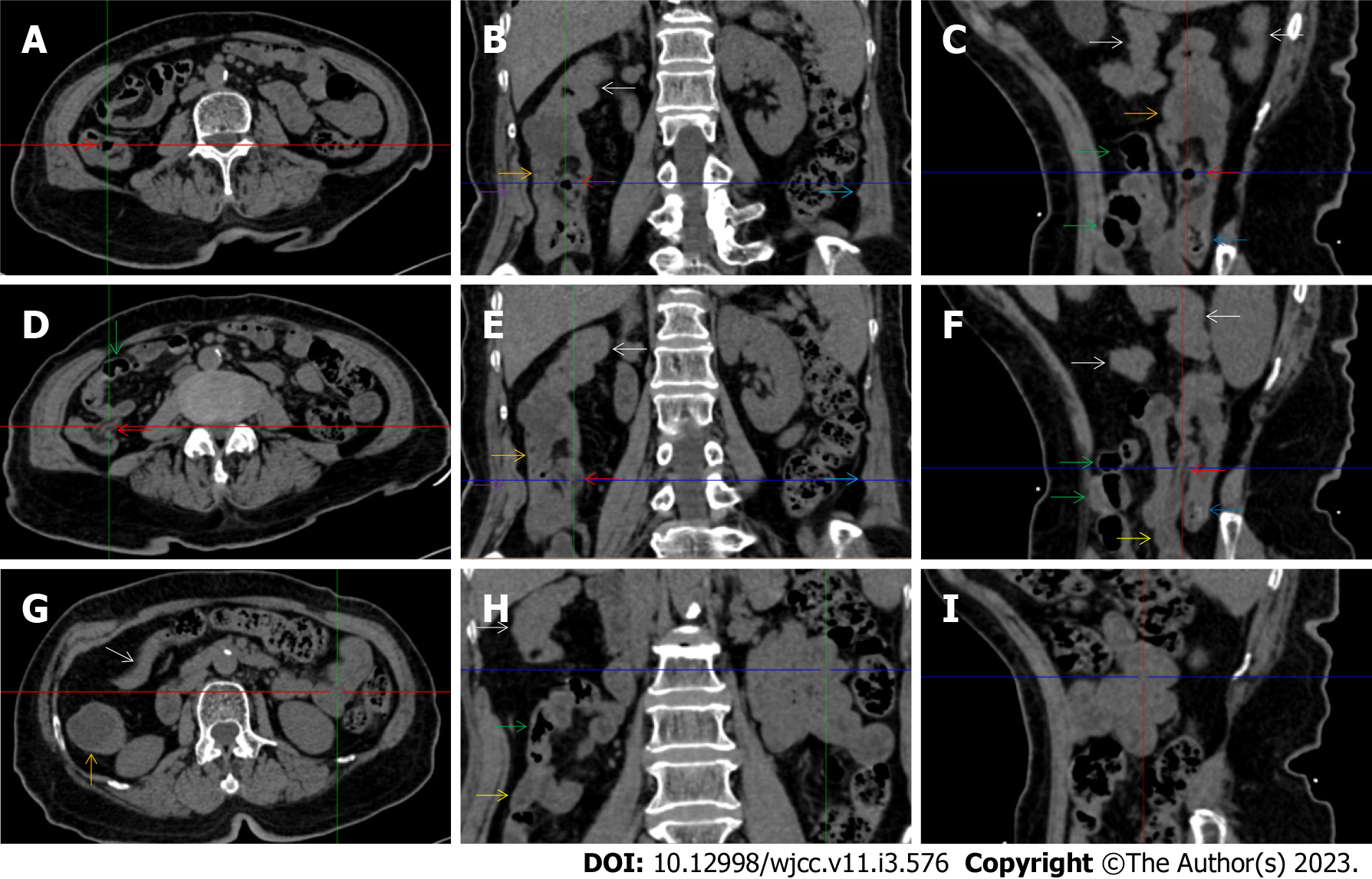
Figure 9 Characteristic images of case 13.
A-C: Characteristic images of the ileocecal valve and ascending colon. There was edematous fat deposition around the ileocecal valve and the terminal ileum, forming the so-called “fat holo sign”. A massive necrotic lesion in the colonic wall was adjacent to the edematous fat deposition, and the serosal colonic wall was hypertrophically thickened (orange arrows). The adjacent parietal peritoneum (purple arrows) and the left parietal peritoneum (navy blue arrows) were also hypertrophically thickened. The mucosa of the cecum was fibrotically thickened (blue arrow); D-F: Characteristic images of the distal ileum. Strictured thickening of the ileocecal valve and terminal ileum (red arrows) led to the distal ileum being liquid-filled. Proximal to the liquid-filled distal ileum (yellow arrows) was the asymmetrically wall-thickened and gas-filled ileum (green arrows). Heterogeneity of the small bowel wall was observed; G-I: Characteristic images of an “abdominal cocoon”. A segment of adhesive bowel loop was present in the middle jejunum, together with the fibrotically thickened bowel and peritoneal involvement forming the so-called “cauliflower sign”. Edematously thickened colonic wall and emptied colonic lumen were present from the proximal ascending colon to the hepatic flexure, followed by a segment of the collapsed transverse colon (white arrows), forming the so-called “empty colon sign”.
- Citation: Zhao XC, Xue CJ, Song H, Gao BH, Han FS, Xiao SX. Bowel inflammatory presentations on computed tomography in adult patients with severe aplastic anemia during flared inflammatory episodes. World J Clin Cases 2023; 11(3): 576-597
- URL: https://www.wjgnet.com/2307-8960/full/v11/i3/576.htm
- DOI: https://dx.doi.org/10.12998/wjcc.v11.i3.576


