Copyright
©The Author(s) 2023.
World J Clin Cases. Jan 26, 2023; 11(3): 576-597
Published online Jan 26, 2023. doi: 10.12998/wjcc.v11.i3.576
Published online Jan 26, 2023. doi: 10.12998/wjcc.v11.i3.576
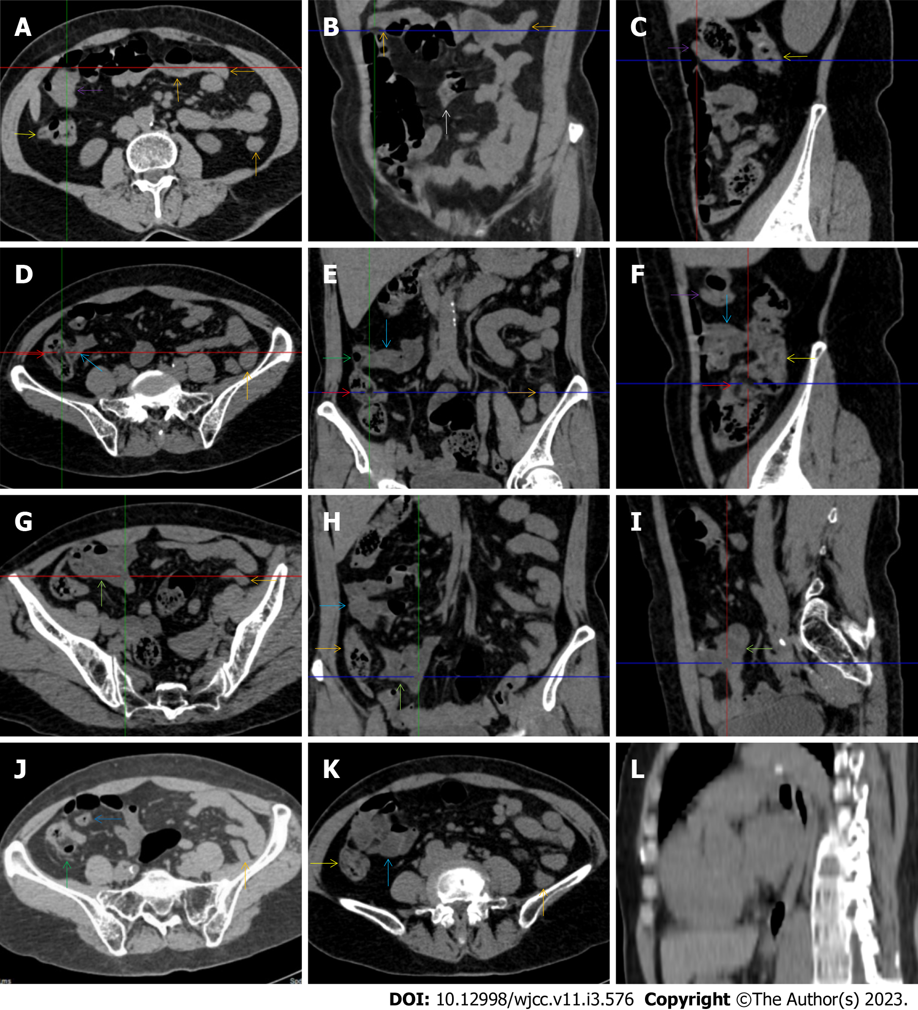
Figure 4 Characteristic images of case 9.
A-C: Characteristic images of an empty colon sign. The segmentally wall-thickened, stratified and emptied colon in the hepatic flexure (purple arrows) followed by the collapsed transverse (adjacent to the dilated ileum in which gas-liquid levels could be recognized) and descending colon (orange arrows), forming the so-called “empty colon sign”. A short segment of asymmetrically thickened wall was present in the proximal ileum (a white arrow), around which the hypervascular fat stranding was especially prominent, distal to which the ileal lumen was gas-filled, and proximal to which the ileal lumen was liquid-filled; D-F: Characteristic images of the ileocecal region. The ileocecal valve and the terminal ileum were thickened and stratified by submucosal fat deposition (red arrows). Omental thickening was especially prominent in the ileocecal region. The wall of the ascending colon was thickened and, in some segments, stratified with submucosal fat deposition, and in other segments, stratified with submucosal edematous tissue (yellow arrows). Several inflamed diverticula (green arrows) were present in the cecum and ascending colon. The distal ileum was strictured (blue arrows), proximal to which the ileal lumen was liquid-filled; G-I: Characteristic images of adhesive bowel loops. The fibrotic mucosa and liquid-filled lumen of the adhesive bowel loops were present in the proximal ileum (powder blue arrows) and distal ileum (jade-green arrows); J: Characteristic image of a balloon sign. A large cluster of circumferentially distributed hypervascular fat stranding wrapped a segment of the dilated lumen and paper-thin bowel wall of the sigmoid colon, forming the so-called “balloon sign”. An inflamed diverticula was present in the cecum, with strikingly thickened omentim (a green arrow); K: Characteristic image of an adhesive bowel loop in the proximal ileum. A segment of adhesive bowel loop in the proximal ileum, together with the fibrotically thickened peritoneum, formed the so-called “abdominal cocoon”; L: Characteristic image of esophagus. Hypertrophic lesions presented in two segments of the esophagus, together with the inflammatory lesions in the jejunum suggesting that the initiating factor in the upper gastrointestinal tract affected the functions of the downstream intestinal segments.
- Citation: Zhao XC, Xue CJ, Song H, Gao BH, Han FS, Xiao SX. Bowel inflammatory presentations on computed tomography in adult patients with severe aplastic anemia during flared inflammatory episodes. World J Clin Cases 2023; 11(3): 576-597
- URL: https://www.wjgnet.com/2307-8960/full/v11/i3/576.htm
- DOI: https://dx.doi.org/10.12998/wjcc.v11.i3.576


