Copyright
©The Author(s) 2023.
World J Clin Cases. Jan 26, 2023; 11(3): 576-597
Published online Jan 26, 2023. doi: 10.12998/wjcc.v11.i3.576
Published online Jan 26, 2023. doi: 10.12998/wjcc.v11.i3.576
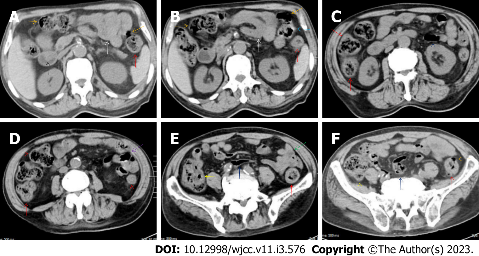
Figure 3 Characteristic images of case 6.
A and B: Characteristic images of the upper abdomen. Several inflamed diverticula (orange arrows) were present in the colonic segments. In the duodenum-jejunum junction (a blue arrow), the bowel wall was fibrotically thickened and the lumen was gas-filled. In the bulb part of the duodenum, a polypoid mass (a black arrow) protruded into the lumen. A segment of adhesive jejunal loop was present in the proximal jejunum (white arrows); C and D: Characteristic images of the thickened colon. The wall from the cecum to the descending colon was significantly thickened with mural stratification, intramural gas and pericolonic fat stranding. A segment of adhesive bowel loop was present in the middle jejunum (a purple arrow) in which the bowel wall was asymmetrically thickened and the lumen was gas-filled with particularly prominent mesenteric fat stranding; E: Characteristic images of the ileocecal region. Stratified thickening of the ileocecal valve and the terminal ileum was gas-filled (yellow arrows). The third segments of adhesive bowel loop were present in the distal jejunum (a green arrow); F: Characteristic image of a balloon sign. The sigmoid colon was dilated and the wall was paper-thin (navy blue arrows), with clustering pericolonic fat stranding forming the so-called “balloon sign”.
- Citation: Zhao XC, Xue CJ, Song H, Gao BH, Han FS, Xiao SX. Bowel inflammatory presentations on computed tomography in adult patients with severe aplastic anemia during flared inflammatory episodes. World J Clin Cases 2023; 11(3): 576-597
- URL: https://www.wjgnet.com/2307-8960/full/v11/i3/576.htm
- DOI: https://dx.doi.org/10.12998/wjcc.v11.i3.576


