Copyright
©The Author(s) 2023.
World J Clin Cases. Oct 16, 2023; 11(29): 7242-7247
Published online Oct 16, 2023. doi: 10.12998/wjcc.v11.i29.7242
Published online Oct 16, 2023. doi: 10.12998/wjcc.v11.i29.7242
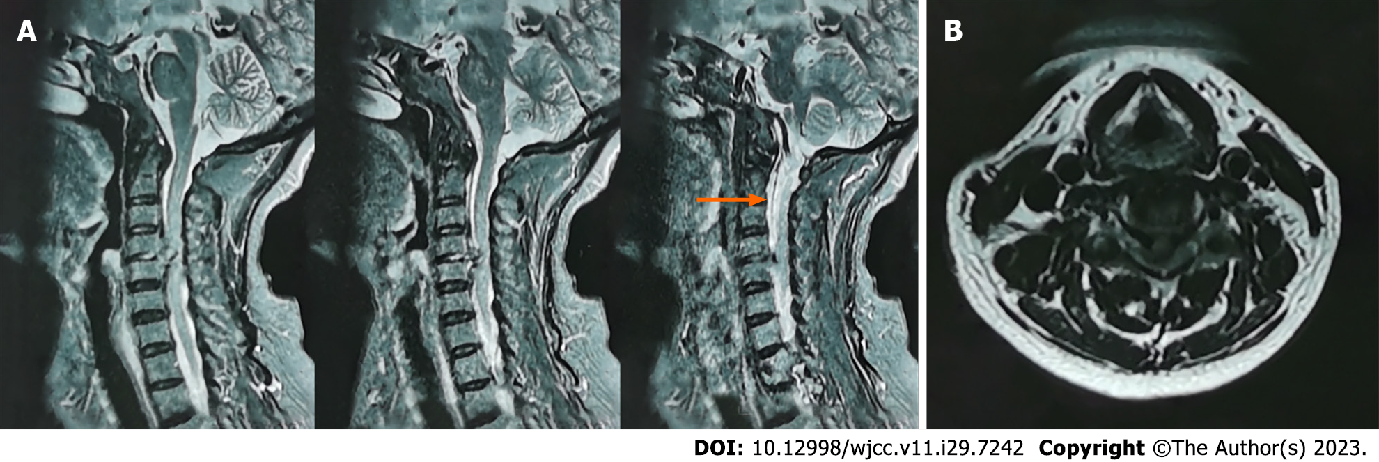
Figure 1 Magnetic resonance imaging of the cervical spine before surgery.
A: Orange arrow shows a strip of leaking cerebrospinal fluid posterior to the C1-4 vertebra; B: Protrusion of the C4/5 disc into the spinal canal, narrowing of the spinal canal at the level of the corresponding disc, loss of the epidural space, compression of the spinal cord, and a high signal area in the medulla.
- Citation: Yu Z, Zhang HFZ, Wang YJ. Surgical treatment of mixed cervical spondylosis with spontaneous cerebrospinal fluid leakage: A case report. World J Clin Cases 2023; 11(29): 7242-7247
- URL: https://www.wjgnet.com/2307-8960/full/v11/i29/7242.htm
- DOI: https://dx.doi.org/10.12998/wjcc.v11.i29.7242


