Copyright
©The Author(s) 2023.
World J Clin Cases. Oct 16, 2023; 11(29): 7234-7241
Published online Oct 16, 2023. doi: 10.12998/wjcc.v11.i29.7234
Published online Oct 16, 2023. doi: 10.12998/wjcc.v11.i29.7234
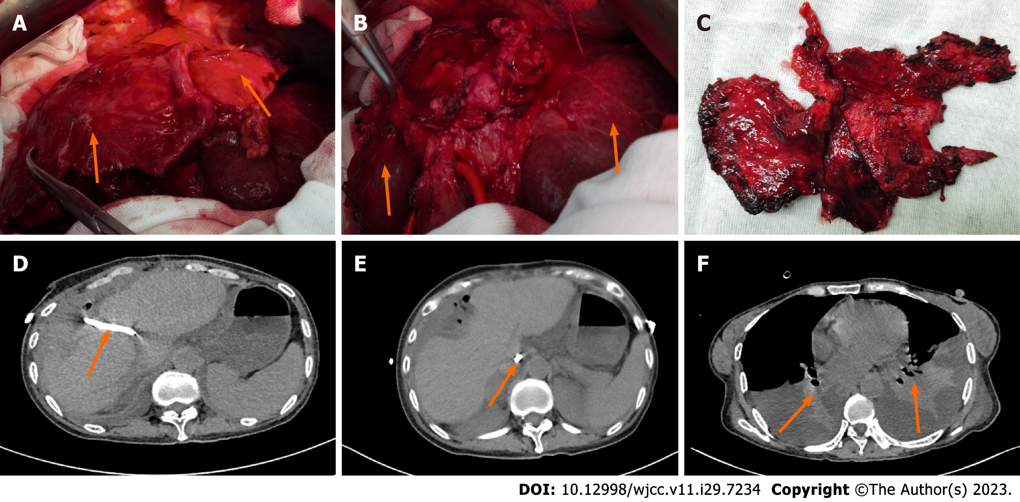
Figure 3 Intraoperative and postoperative images.
A: Obvious atrophy and thinning of the left and right liver lobes were observed during the operation; B: After resection of the left and right liver lobes, the caudate lobe of the liver with huge hyperplasia was shown, as indicated by the green arrows; C: Left and right liver specimens; D: The arrow indicates percutaneous transhepatic placement of a drainage tube into the dilated intrahepatic bile duct; E: The tip of the drainage tube was inserted into the abdominal cavity, as indicated by the arrow, a small amount of fluid was collected in the abdominal cavity; F: Atelectasis of both lower lobes accompanied by pleural effusion.
- Citation: Liang SY, Lu JG, Wang ZD. Imaging misdiagnosis and clinical analysis of significant hepatic atrophy after bilioenteric anastomosis: A case report. World J Clin Cases 2023; 11(29): 7234-7241
- URL: https://www.wjgnet.com/2307-8960/full/v11/i29/7234.htm
- DOI: https://dx.doi.org/10.12998/wjcc.v11.i29.7234


