Copyright
©The Author(s) 2023.
World J Clin Cases. Oct 16, 2023; 11(29): 7207-7213
Published online Oct 16, 2023. doi: 10.12998/wjcc.v11.i29.7207
Published online Oct 16, 2023. doi: 10.12998/wjcc.v11.i29.7207
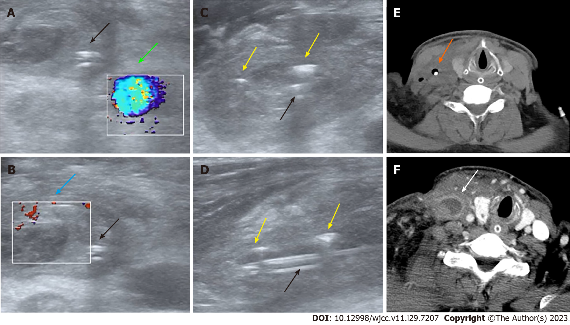
Figure 1 Ultrasound and computed tomography images of emphysematous thrombophlebitis caused by a misplaced central venous catheter.
A and B: Panel A and B: Color Doppler ultrasound on day 9 after central venous catheter (CVC) insertion showing normal blood flow in the right internal carotid artery (green arrow) and thrombosis in the dilated right internal jugular vein (IJV) without blood flow (blue arrow) (transverse section); C and D: Transverse section (C) and longitudinal section (D). Ultrasound on day 9 after CVC insertion showing the misplaced CVC (black arrows) and gas bubbles in the IJV manifesting as hyperechoic lines with dirty shadowing and comet-tail artifacts (yellow arrows); E: Computed tomography (CT) image on day 9 after CVC insertion showing air bubbles surrounding the misplaced CVC (orange arrow) (transverse section); F: Contrast-enhanced CT image on day 2 after CVC removal showing the swollen right IJV filled with thrombus material but no gas (white arrow) (transverse section).
- Citation: Chen N, Chen HJ, Chen T, Zhang W, Fu XY, Xing ZX. Emphysematous thrombophlebitis caused by a misplaced central venous catheter: A case report. World J Clin Cases 2023; 11(29): 7207-7213
- URL: https://www.wjgnet.com/2307-8960/full/v11/i29/7207.htm
- DOI: https://dx.doi.org/10.12998/wjcc.v11.i29.7207


