Copyright
©The Author(s) 2023.
World J Clin Cases. Oct 16, 2023; 11(29): 7162-7169
Published online Oct 16, 2023. doi: 10.12998/wjcc.v11.i29.7162
Published online Oct 16, 2023. doi: 10.12998/wjcc.v11.i29.7162
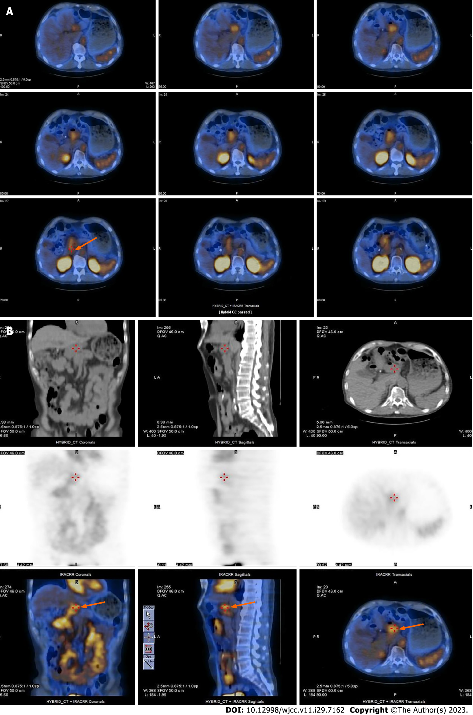
Figure 3 Single-photon emission computed tomography/computed tomography localized a bleeding point at duodenum level.
A: Single-photon emission computed tomography/computed tomography (SPECT/CT) transverse images (orange arrow); B: Top was CT three-dimensional (3D) images, middle was SPECT 3D images, and bottom was SPECT/CT 3D images (orange arrow).
- Citation: Kuo CL, Chen CF, Su WK, Yang RH, Chang YH. Rare finding of primary aortoduodenal fistula on single-photon emission computed tomography/computed tomography of gastrointestinal bleeding: A case report. World J Clin Cases 2023; 11(29): 7162-7169
- URL: https://www.wjgnet.com/2307-8960/full/v11/i29/7162.htm
- DOI: https://dx.doi.org/10.12998/wjcc.v11.i29.7162


