Copyright
©The Author(s) 2023.
World J Clin Cases. Oct 16, 2023; 11(29): 7127-7135
Published online Oct 16, 2023. doi: 10.12998/wjcc.v11.i29.7127
Published online Oct 16, 2023. doi: 10.12998/wjcc.v11.i29.7127
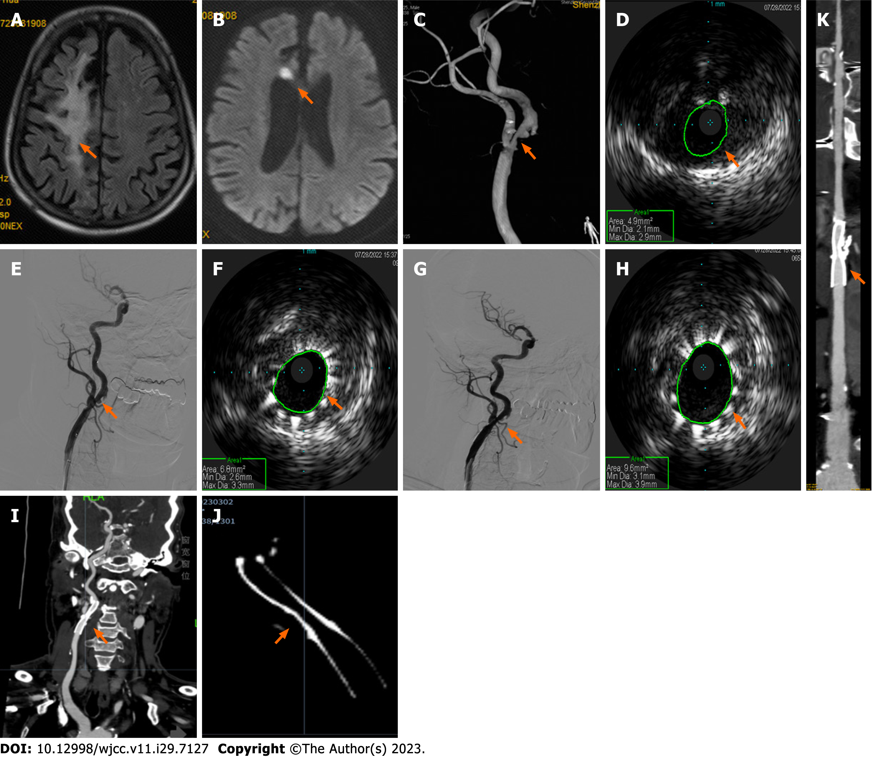
Figure 1 Neuroimaging results of patient one during intravascular ultrasound-assisted carotid artery stenting.
A and B: Brain magnetic resonance imaging demonstrates a paraventricular white-matter lesion in the right frontal lobe (A, arrow) on fluid-attenuated inversion recovery imaging, and a small acute infarction at the genu of the corpus callosum on diffusion weighted imaging (B, arrow); C: Angiography shows 70% stenosis of the initial segment of the right internal carotid artery (ICA) (arrow); D and E: Preoperative intravascular ultrasound (IVUS) displays severe stenosis with plaque formation and obvious calcification under plaque (D, arrow) and a well-positioned stent (E, arrow); F-H: Subsequent IVUS imaging confirmed improvement of the narrowest part (F, arrow), thus post-stent balloon dilatation was performed. Postoperative angiography shows a right ICA residual stenosis of 20% (G, arrow), and IVUS confirmed good stent expansion and adherence (H, arrow); I-K: Computed tomography angiography six months postoperatively revealed mild in-stent stenosis in the right initial segment of the ICA (I, multiplanar reconstruction; J, amplification imaging with adjusting parameters; K, curve planar reformation; arrows).
- Citation: Fu PC, Wang JY, Su Y, Liao YQ, Li SL, Xu GL, Huang YJ, Hu MH, Cao LM. Intravascular ultrasonography assisted carotid artery stenting for treatment of carotid stenosis: Two case reports. World J Clin Cases 2023; 11(29): 7127-7135
- URL: https://www.wjgnet.com/2307-8960/full/v11/i29/7127.htm
- DOI: https://dx.doi.org/10.12998/wjcc.v11.i29.7127


