Copyright
©The Author(s) 2023.
World J Clin Cases. Oct 16, 2023; 11(29): 7113-7126
Published online Oct 16, 2023. doi: 10.12998/wjcc.v11.i29.7113
Published online Oct 16, 2023. doi: 10.12998/wjcc.v11.i29.7113
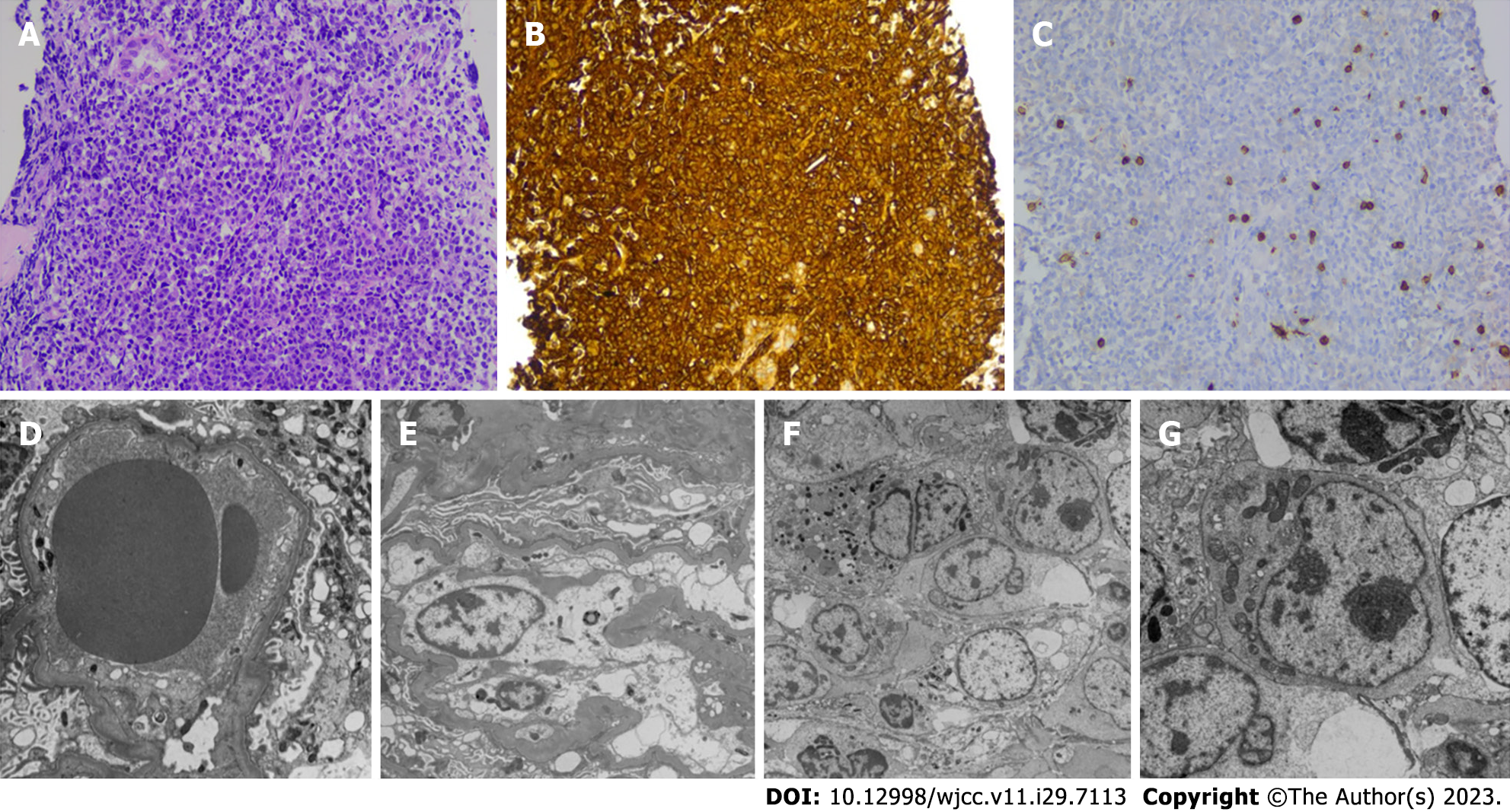
Figure 1 Histologic, immunohistochemical examination and electron-microscopic findings.
A: Diffuse infiltration of pleomorphic cells is identified throughout the specimen (HE × 5) (× 2.0 k); B: The cells were immunoreactive for CD20 (× 20) (× 1.5 k); C: Negative for CD3 (× 20) (× 1.0 k); D and E: No electron-dense deposit is recognized, and glomerular basement membrane appeared normal in thickness, contour, and texture; however, strikingly, diffuse prominent infiltration of atypical lymphocytes is seen in the interstitium (× 2.0 k); F and G: The cells exhibited round to oval cleaved and non-cleaved nuclei with variable clumping of chromatin and large prominent marginated nucleoli.
- Citation: Lee SB, Yoon YM, Hong R. Primary renal lymphoma presenting as renal failure: A case report and review of literature from 1989. World J Clin Cases 2023; 11(29): 7113-7126
- URL: https://www.wjgnet.com/2307-8960/full/v11/i29/7113.htm
- DOI: https://dx.doi.org/10.12998/wjcc.v11.i29.7113


