Copyright
©The Author(s) 2023.
World J Clin Cases. Oct 6, 2023; 11(28): 6967-6973
Published online Oct 6, 2023. doi: 10.12998/wjcc.v11.i28.6967
Published online Oct 6, 2023. doi: 10.12998/wjcc.v11.i28.6967
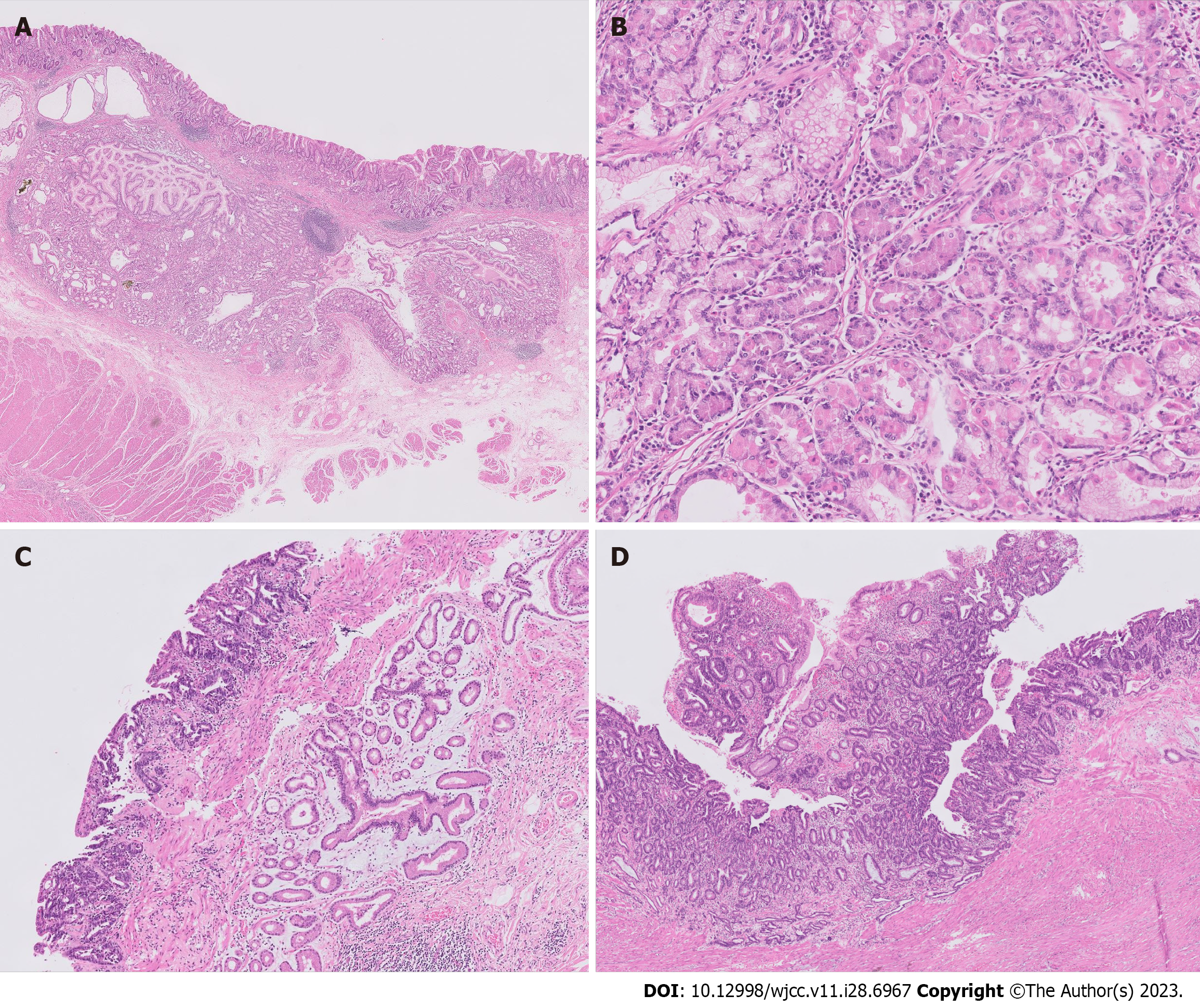
Figure 3 Microscopic findings of the surgical specimen (hematoxylin and eosin staining).
A: Gastric hamartomatous inverted polyp (GHIP) is located in the submucosal layer with focal cystic dilations and is surrounded by smooth muscle bundles. On the left upper side, gastritis cystica profundas is adjacent to the GHIP (20 × magnification); B: GHIP consists of heterogeneous glandular cell types (100 × magnification); C: GHIP is positioned separately under a well differentiated adenocarcinoma (40 × magnification); D: Adenocarcinoma extending to the muscularis propria is shown (40 × magnification).
- Citation: Park G, Kim J, Lee SH, Kim Y. Large gastric hamartomatous inverted polyp accompanied by advanced gastric cancer: A case report. World J Clin Cases 2023; 11(28): 6967-6973
- URL: https://www.wjgnet.com/2307-8960/full/v11/i28/6967.htm
- DOI: https://dx.doi.org/10.12998/wjcc.v11.i28.6967


