Copyright
©The Author(s) 2023.
World J Clin Cases. Oct 6, 2023; 11(28): 6967-6973
Published online Oct 6, 2023. doi: 10.12998/wjcc.v11.i28.6967
Published online Oct 6, 2023. doi: 10.12998/wjcc.v11.i28.6967
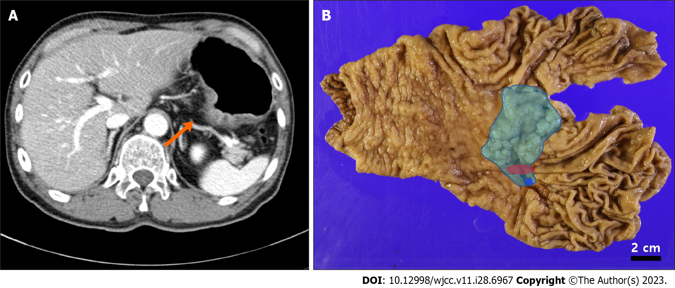
Figure 2 Preoperative image study and gross lesions of the stomach.
A: Preoperative abdominal computed tomography detected a 4.6 cm-sized lesion at the posterior wall of the mid-body (orange arrow); B: On the total gastrectomy specimen, a 6.9 cm × 4.5 cm-sized ulcero-infiltrative gastric hamartomatous inverted polyps extending from the lesser curvature of the cardia to the posterior wall of the body (blue) and a 1.6 cm-sized adenocarcinoma located at the posterior wall of the body (red) were indicated. The two lesions showed an overlap.
- Citation: Park G, Kim J, Lee SH, Kim Y. Large gastric hamartomatous inverted polyp accompanied by advanced gastric cancer: A case report. World J Clin Cases 2023; 11(28): 6967-6973
- URL: https://www.wjgnet.com/2307-8960/full/v11/i28/6967.htm
- DOI: https://dx.doi.org/10.12998/wjcc.v11.i28.6967


