Copyright
©The Author(s) 2023.
World J Clin Cases. Oct 6, 2023; 11(28): 6967-6973
Published online Oct 6, 2023. doi: 10.12998/wjcc.v11.i28.6967
Published online Oct 6, 2023. doi: 10.12998/wjcc.v11.i28.6967
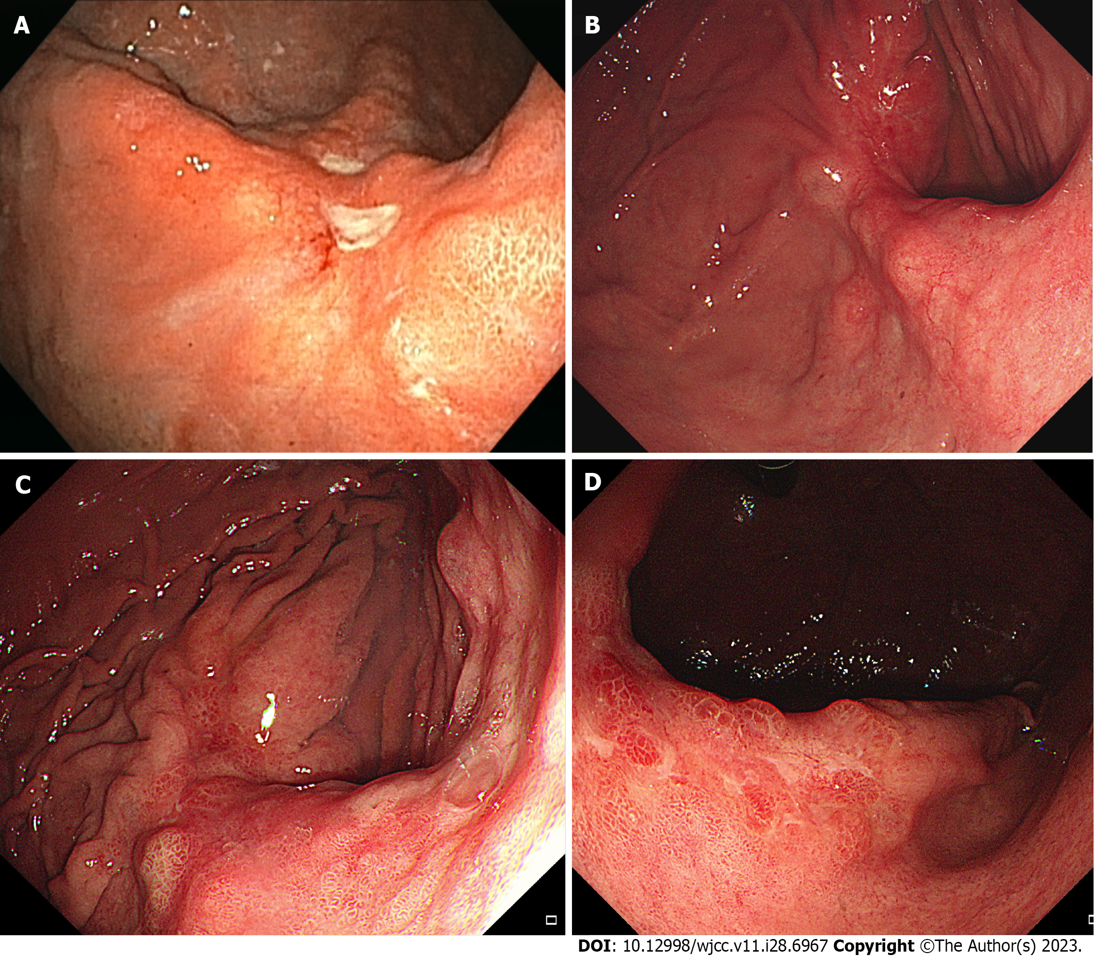
Figure 1 Initial and follow-up endoscopic findings.
A: Initial esophagogastroduodenoscopy (EGD) showing two whitish ulcerative lesions at the posterior wall of the mid-body; B: In the first follow-up EGD, the lesions are healed with a reddish scar and several bulging areas of normal mucosa on the peripheral; C: The last follow-up EGD presents a 4.5 cm-sized ulcero-infiltrative lesion in the posterior wall of the mid-body; D: In the retroflexed view, this lesion is covered with an unevenly distributed regenerative epithelium and whitish discoloration. The tumor is identified as a well-differentiated adenocarcinoma.
- Citation: Park G, Kim J, Lee SH, Kim Y. Large gastric hamartomatous inverted polyp accompanied by advanced gastric cancer: A case report. World J Clin Cases 2023; 11(28): 6967-6973
- URL: https://www.wjgnet.com/2307-8960/full/v11/i28/6967.htm
- DOI: https://dx.doi.org/10.12998/wjcc.v11.i28.6967


