Copyright
©The Author(s) 2023.
World J Clin Cases. Oct 6, 2023; 11(28): 6908-6919
Published online Oct 6, 2023. doi: 10.12998/wjcc.v11.i28.6908
Published online Oct 6, 2023. doi: 10.12998/wjcc.v11.i28.6908
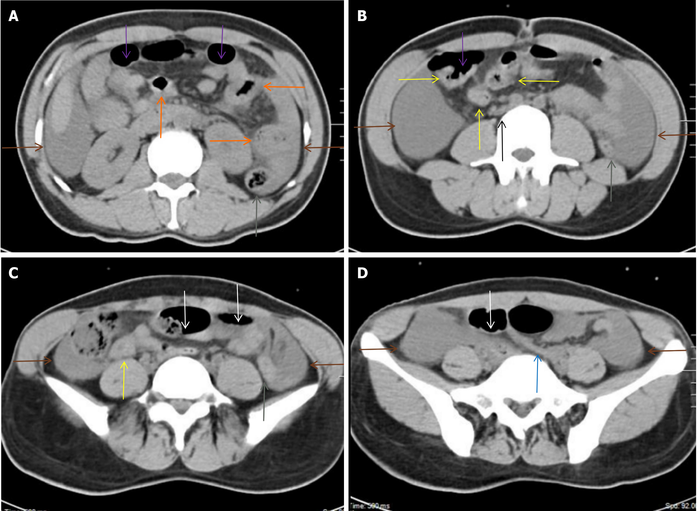
Figure 3 Abdominal computed tomography scan during active tuberculosis.
A: From the duodenum to the proximal ileum, the bowel wall was segmentally thickened, with perienteric inflammatory changes (orange arrows). Perienteric fat stranding was especially striking adjacent to the homogeneously thickened walls and gas-filled lumen of several segments of the small intestine; B and C: The homogeneously thickened walls of the distal ileum and the cecum (yellow arrows) were surrounded by a cluster of misty fat stranding in the right iliac fossa, with adjacent lymphadenopathy (a black arrow). The ascending and transverse colon were dilated (purple arrows), whereas most of the descending and proximal sigmoid colon were collapsed (gray arrows). However, the middle sigmoid colon was dilated (white arrows). Moderate ascites was present in the peritoneal cavity (brown arrows), which indicated peritoneal involvement; D: Bowel wall thickening with a collapsed colonic lumen was also present in the distal sigmoid colon (blue arrow). These imaging features suggested that tuberculosis infected the intestines and peritoneum.
- Citation: Xiu NN, Yang XD, Xu J, Ju B, Sun XY, Zhao XC. Leukemic transformation during anti-tuberculosis treatment in aplastic anemia-paroxysmal nocturnal hemoglobinuria syndrome: A case report and review of literature. World J Clin Cases 2023; 11(28): 6908-6919
- URL: https://www.wjgnet.com/2307-8960/full/v11/i28/6908.htm
- DOI: https://dx.doi.org/10.12998/wjcc.v11.i28.6908


