Copyright
©The Author(s) 2023.
World J Clin Cases. Oct 6, 2023; 11(28): 6889-6894
Published online Oct 6, 2023. doi: 10.12998/wjcc.v11.i28.6889
Published online Oct 6, 2023. doi: 10.12998/wjcc.v11.i28.6889
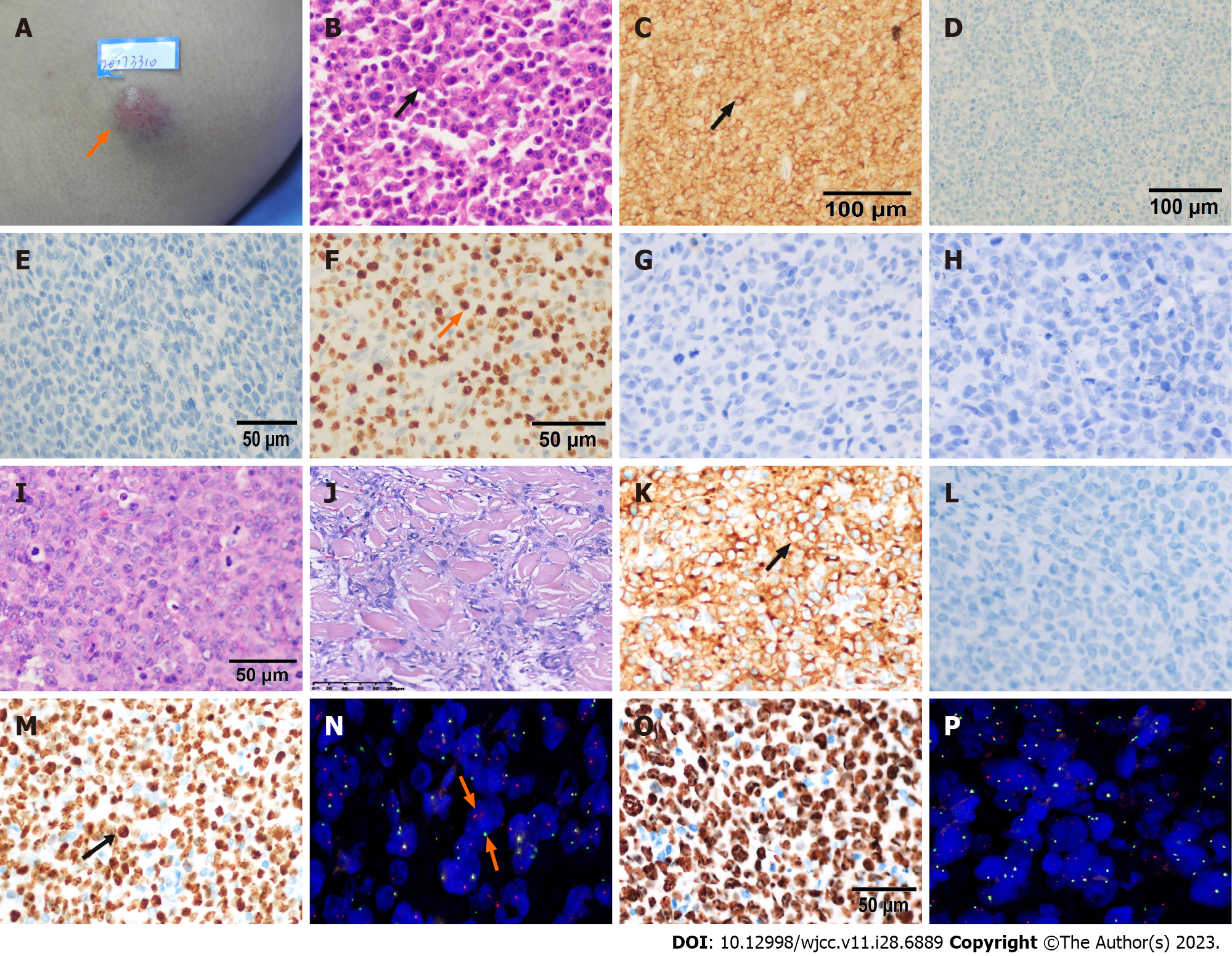
Figure 1 Morphologic and immunohistochemical characterization of this patient’s primary cutaneous anaplastic large cell lymphoma (A-H) and the markers at two times of his relapses (I-N for first relapse, O-P for second relapse).
Unless otherwise indicated, the images are taken using 400× magnification. A: A 2 cm × 2 cm cutaneous swelling at left upper arm with many capillaries on the surface (arrow); B: Anaplastic large cell lymphoma with diffuse, cohesive sheets of enlarged cells with anaplastic morphology by hematoxylin and eosin (HE) (arrow); C: CD30 was strong, diffusely positive (80%) on the membrane of tumor cells (arrow), ×200; D: ALK was nagative in both cell nucleus and cytoplasma, ×200; E: EMA was cytoplasmic and membranaous nagative; F: Ki-67 was positive in 80% nucleoplasm and nucleoli rim (arrow). G: CD20 was negative. H: PAX5 was negative. I: Diffuse, cohesive lymphoma cells with anaplastic, pleomorphic appearance when the patient first relapsed by HE; J: Anaplastic large cell lymphoma tumor cells infiltrating in muscle layer; K: CD30 was difuse membranous positive (arrow); L: Anaplastic lymphoma kinase staining was negative; M: Ki67 was 95% in nucleus positive; N: Interferon regulatory factor 4 (IRF4) breakage was detected in 80% tumor cells by immunofluorescence hybridization (arrow). Spectrum Red stands for 5’ IRF4, Spectrum Green stands for 3’ IRF4; O: Ki67 was 90% positive when this patient relapsed secondly; P: IRF4 breakage was positive in 70% tumor cells.
- Citation: Mu HX, Tang XQ. Primary cutaneous anaplastic large cell lymphoma with over-expressed Ki-67 transitioning into systemic anaplastic large cell lymphoma: A case report. World J Clin Cases 2023; 11(28): 6889-6894
- URL: https://www.wjgnet.com/2307-8960/full/v11/i28/6889.htm
- DOI: https://dx.doi.org/10.12998/wjcc.v11.i28.6889


