Copyright
©The Author(s) 2023.
World J Clin Cases. Oct 6, 2023; 11(28): 6831-6840
Published online Oct 6, 2023. doi: 10.12998/wjcc.v11.i28.6831
Published online Oct 6, 2023. doi: 10.12998/wjcc.v11.i28.6831
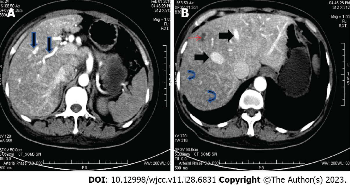
Figure 2 Computed tomography scan of the abdomen in the patient with hereditary hemorrhagic telangiectasia.
A: Dilated and tortuous hepatic arteries (arrow); B: Early enhancement of enlarged hepatic veins (thick arrows), diffuse parenchymal telangiectasia (curved arrows), and an incidentally found liver cyst (thin arrow).
- Citation: Chen YL, Jiang HY, Li DP, Lin J, Chen Y, Xu LL, Gao H. Multi-organ hereditary hemorrhagic telangiectasia: A case report. World J Clin Cases 2023; 11(28): 6831-6840
- URL: https://www.wjgnet.com/2307-8960/full/v11/i28/6831.htm
- DOI: https://dx.doi.org/10.12998/wjcc.v11.i28.6831


