Copyright
©The Author(s) 2023.
World J Clin Cases. Oct 6, 2023; 11(28): 6797-6805
Published online Oct 6, 2023. doi: 10.12998/wjcc.v11.i28.6797
Published online Oct 6, 2023. doi: 10.12998/wjcc.v11.i28.6797
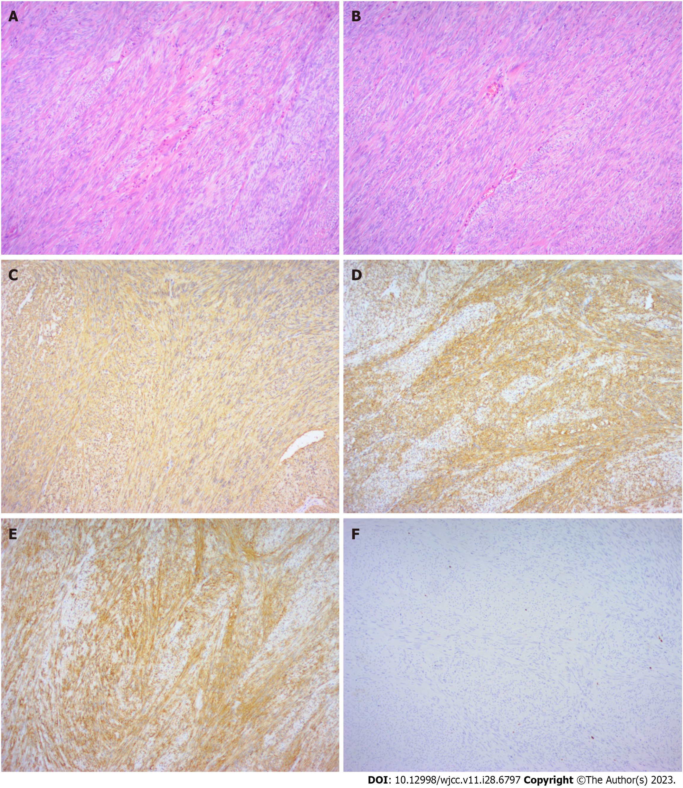
Figure 5 Histopathology of the biopsy specimen.
A and B: Histopathology showed that the cells were spindle-shaped and moderately differentiated (magnifications, × 100). C to F: Immunohistochemistry results showed that cluster of differentiation (CD) 34 (C), CD117 (D), Dog-1 (E), and CD34 (F) were all positive, and Ki67 was negative.
- Citation: Dong RX, Wang C, Zhou H, Yin HQ, Liu Y, Liang HT, Pan YB, Wang JW, Cao YQ. Rare rectal gastrointestinal stromal tumor case: A case report and review of the literature. World J Clin Cases 2023; 11(28): 6797-6805
- URL: https://www.wjgnet.com/2307-8960/full/v11/i28/6797.htm
- DOI: https://dx.doi.org/10.12998/wjcc.v11.i28.6797


