Copyright
©The Author(s) 2023.
World J Clin Cases. Sep 26, 2023; 11(27): 6631-6639
Published online Sep 26, 2023. doi: 10.12998/wjcc.v11.i27.6631
Published online Sep 26, 2023. doi: 10.12998/wjcc.v11.i27.6631
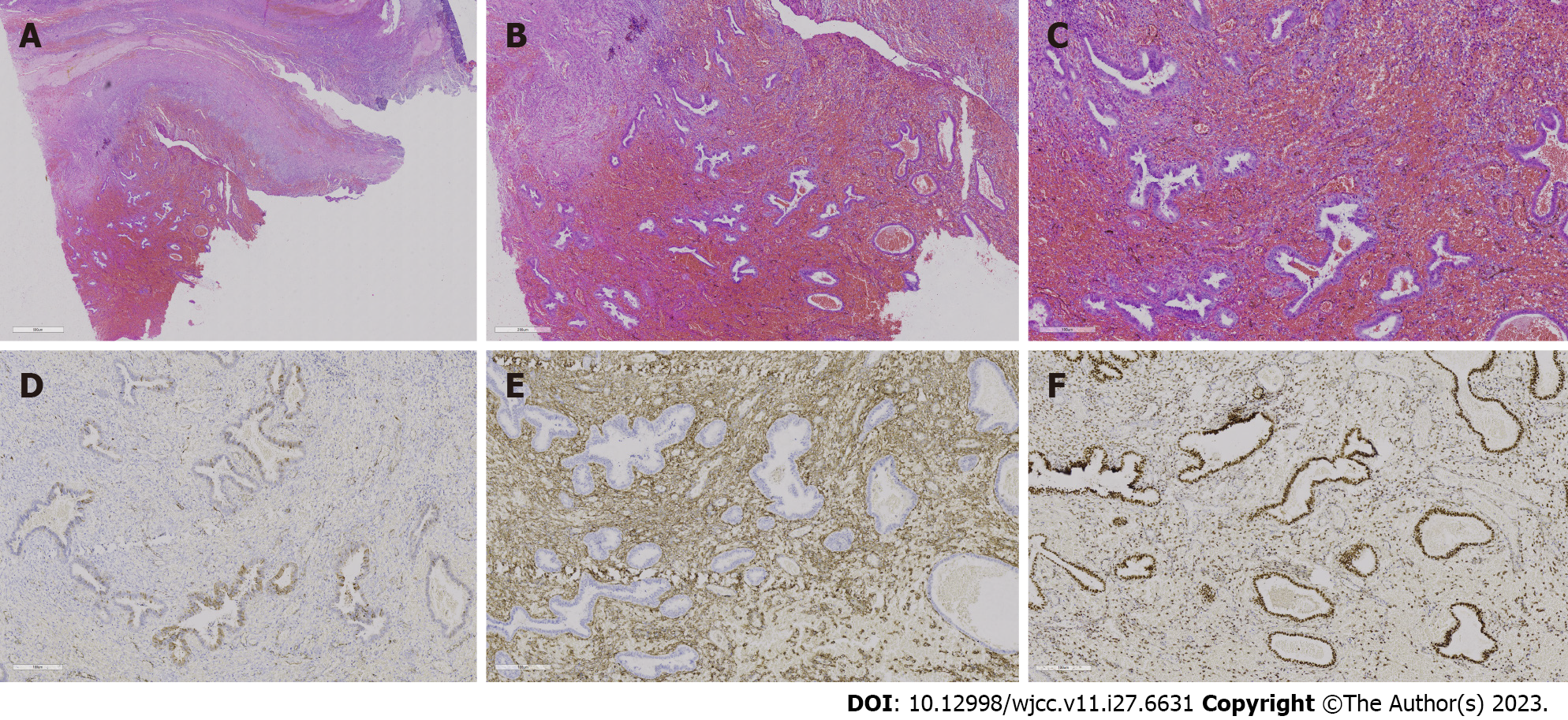
Figure 6 Histopathological analysis and immunohistochemical examination of the resected specimen.
A-C: Hepatic encephalopathy 10 ×, 20 ×, and 40 ×, respectively. Endometrial stroma and glands are observed in the cyst, and extensive haemorrhagic foci are shown without typical thick-walled blood vessels; D: Represents P16, and the visible part of the endometrial glands is positive [immunohistochemistry (IHC) 20 ×]; E: CD10 and endometrial stromal expression is strongly positive, but the glands are negative (IHC 20 ×); F: Represents the oestrogen receptor, with the endometrial glands strongly positive and the stroma partially positive (IHC 20 ×).
- Citation: Zhang DY, Peng C, Huang Y, Cao JC, Zhou YF. Rapidly growing extensive polypoid endometriosis after gonadotropin-releasing hormone agonist discontinuation: A case report. World J Clin Cases 2023; 11(27): 6631-6639
- URL: https://www.wjgnet.com/2307-8960/full/v11/i27/6631.htm
- DOI: https://dx.doi.org/10.12998/wjcc.v11.i27.6631


