Copyright
©The Author(s) 2023.
World J Clin Cases. Sep 26, 2023; 11(27): 6631-6639
Published online Sep 26, 2023. doi: 10.12998/wjcc.v11.i27.6631
Published online Sep 26, 2023. doi: 10.12998/wjcc.v11.i27.6631
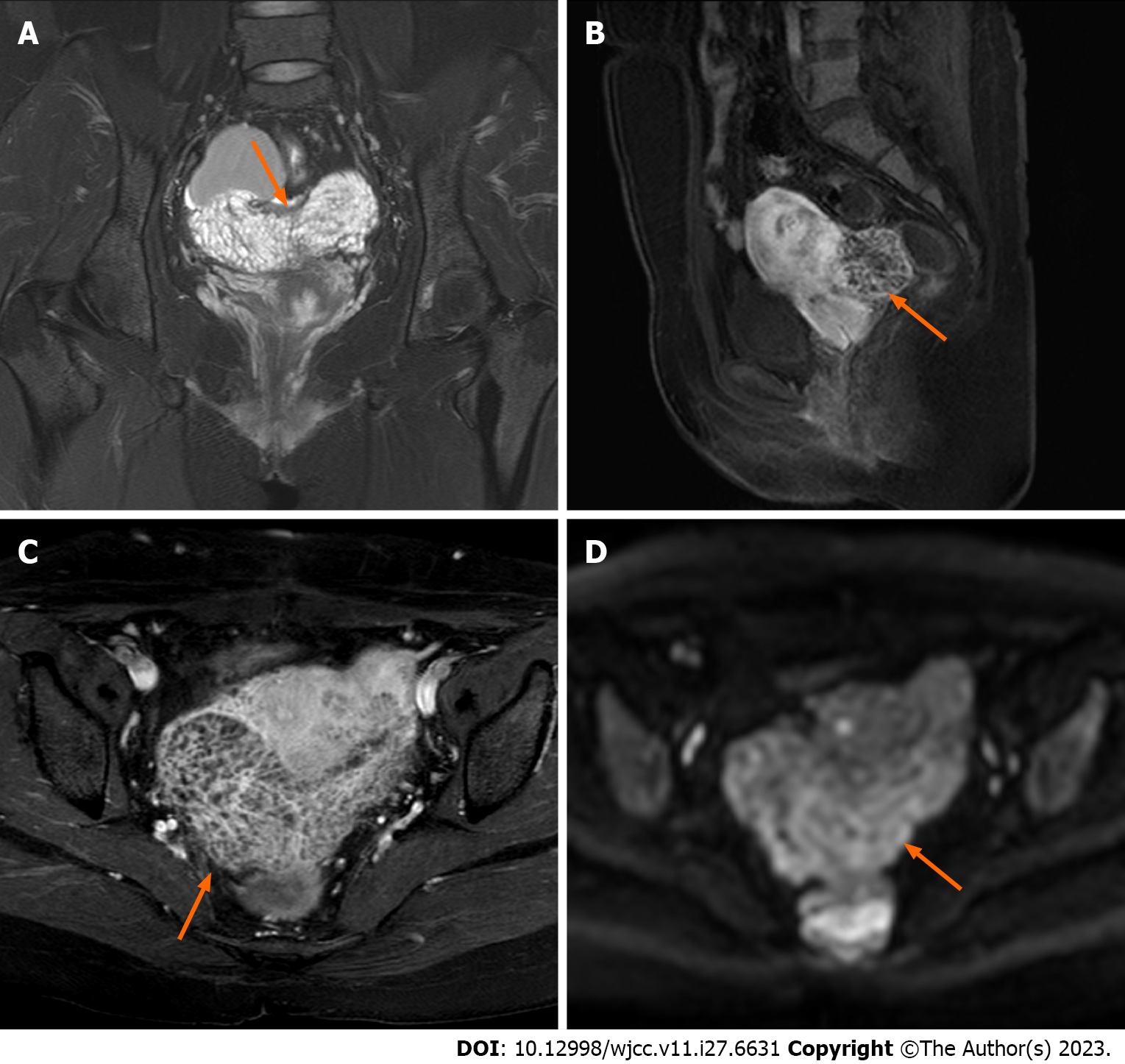
Figure 3 Magnetic resonance imaging scans acquired after one month of cessation of gonadotropin-releasing hormone agonist therapy.
A: In the coronal plane, the lesion displays a subtle elevation in signal intensity on T2-weighted imaging, characterized by diffuse and homogenous fine septal modifications internally (orange arrow); B and C: In the sagittal and axial enhanced scans, the lesion demonstrates a slightly higher enhancement in both the margins and internal septations (orange arrow); D The lesion exhibits a slight increase in the diffusion-weighted imaging signal (orange arrow).
- Citation: Zhang DY, Peng C, Huang Y, Cao JC, Zhou YF. Rapidly growing extensive polypoid endometriosis after gonadotropin-releasing hormone agonist discontinuation: A case report. World J Clin Cases 2023; 11(27): 6631-6639
- URL: https://www.wjgnet.com/2307-8960/full/v11/i27/6631.htm
- DOI: https://dx.doi.org/10.12998/wjcc.v11.i27.6631


