Copyright
©The Author(s) 2023.
World J Clin Cases. Sep 26, 2023; 11(27): 6631-6639
Published online Sep 26, 2023. doi: 10.12998/wjcc.v11.i27.6631
Published online Sep 26, 2023. doi: 10.12998/wjcc.v11.i27.6631
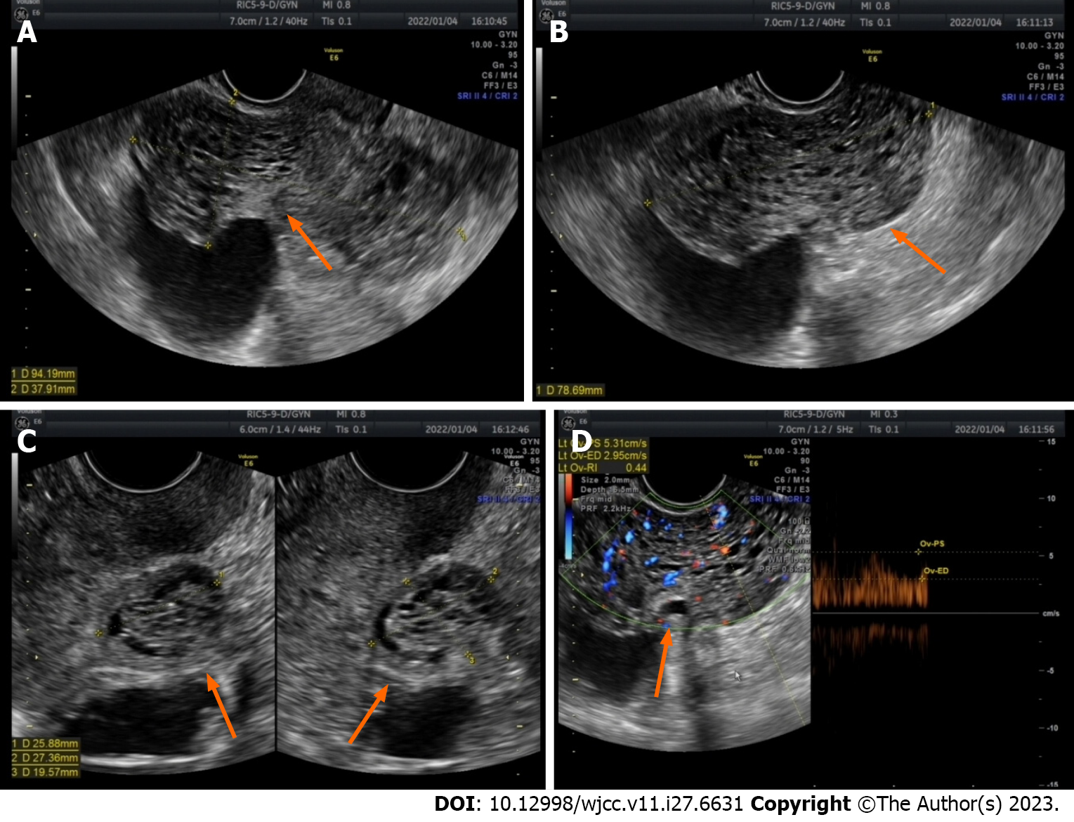
Figure 2 Ultrasound image of the lesion 1 mo after gonadotropin-releasing hormone agonist therapy was discontinued.
A-C: After discontinuation of gonadotropin-releasing hormone agonist for one month, the maximum diameter of the lesion increased to 9.4 cm, with numerous anechoic areas visible inside, exhibiting a honeycomb-like pattern of change (orange arrow); D: Abundant blood flow signals are observed within the lesion, with a blood flow resistance of 0.44 (orange arrow).
- Citation: Zhang DY, Peng C, Huang Y, Cao JC, Zhou YF. Rapidly growing extensive polypoid endometriosis after gonadotropin-releasing hormone agonist discontinuation: A case report. World J Clin Cases 2023; 11(27): 6631-6639
- URL: https://www.wjgnet.com/2307-8960/full/v11/i27/6631.htm
- DOI: https://dx.doi.org/10.12998/wjcc.v11.i27.6631


