Copyright
©The Author(s) 2023.
World J Clin Cases. Sep 26, 2023; 11(27): 6551-6557
Published online Sep 26, 2023. doi: 10.12998/wjcc.v11.i27.6551
Published online Sep 26, 2023. doi: 10.12998/wjcc.v11.i27.6551
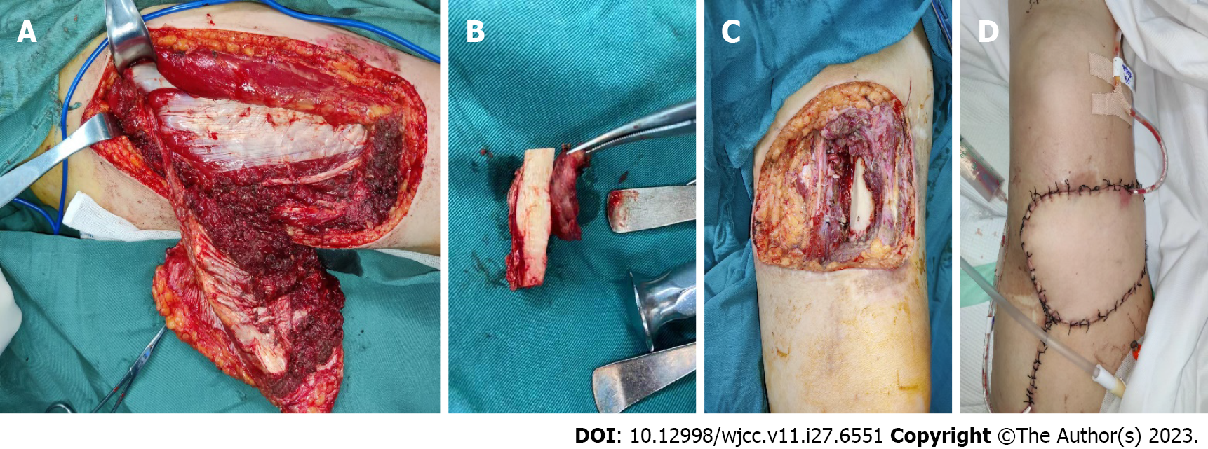
Figure 5 Illustration of patient undergoing surgery on March 3, 2022.
A–D: Surgical images. A: A tissue breaks down (about 8 cm × 10 cm × 5 cm) was seen above the knee joint on the left thigh, deep to the medial, lateral, and rectus femoris muscles; B: Trauma after excision of the mass; C: Left thigh swelling: Gray/yellow/brown soft tissue with skin (10 cm × 6 cm × 3.5 cm) and a gray/white/brown mass (4 cm × 3.5 cm × 2.5 cm) on the skin surface; D: The wound was closed with an 8 cm × 6 cm flap and a drainage tube was left in place.
- Citation: Zhu YQ, Zhao GC, Zheng CX, Yuan L, Yuan GB. Managing spindle cell sarcoma with surgery and high-intensity focused ultrasound: A case report. World J Clin Cases 2023; 11(27): 6551-6557
- URL: https://www.wjgnet.com/2307-8960/full/v11/i27/6551.htm
- DOI: https://dx.doi.org/10.12998/wjcc.v11.i27.6551


