Copyright
©The Author(s) 2023.
World J Clin Cases. Sep 26, 2023; 11(27): 6551-6557
Published online Sep 26, 2023. doi: 10.12998/wjcc.v11.i27.6551
Published online Sep 26, 2023. doi: 10.12998/wjcc.v11.i27.6551
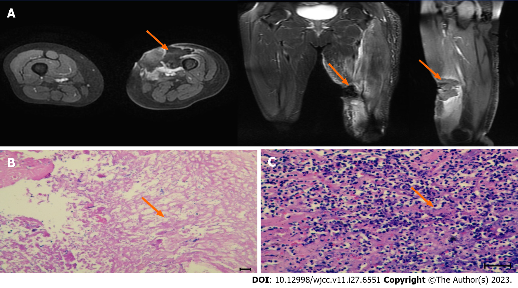
Figure 2 Magnetic resonance imaging imaging and pathological biopsy after the first high-intensity focused ultrasound treatment.
A: An ovoid mass (about 26 mm × 36 mm × 36 mm) with an equal/slightly low signal on T1WI and a slightly low/high mixed signal on T2WI was seen under the skin of the anterior medial part of the left mid-thigh, with a clear border. The lesion was mildly enhanced at the edge after enhancement. A patchy slightly high signal on T2WI with poorly defined borders was seen adjacent to the left middle femur (about 11 mm × 14 mm × 55 mm), with heterogeneous enhancement after enhancement; B and C: H&E staining showing acute and chronic inflammation with necrotic granulomatous tissue proliferation. Scale bar: 100 μm.
- Citation: Zhu YQ, Zhao GC, Zheng CX, Yuan L, Yuan GB. Managing spindle cell sarcoma with surgery and high-intensity focused ultrasound: A case report. World J Clin Cases 2023; 11(27): 6551-6557
- URL: https://www.wjgnet.com/2307-8960/full/v11/i27/6551.htm
- DOI: https://dx.doi.org/10.12998/wjcc.v11.i27.6551


