Copyright
©The Author(s) 2023.
World J Clin Cases. Sep 26, 2023; 11(27): 6551-6557
Published online Sep 26, 2023. doi: 10.12998/wjcc.v11.i27.6551
Published online Sep 26, 2023. doi: 10.12998/wjcc.v11.i27.6551
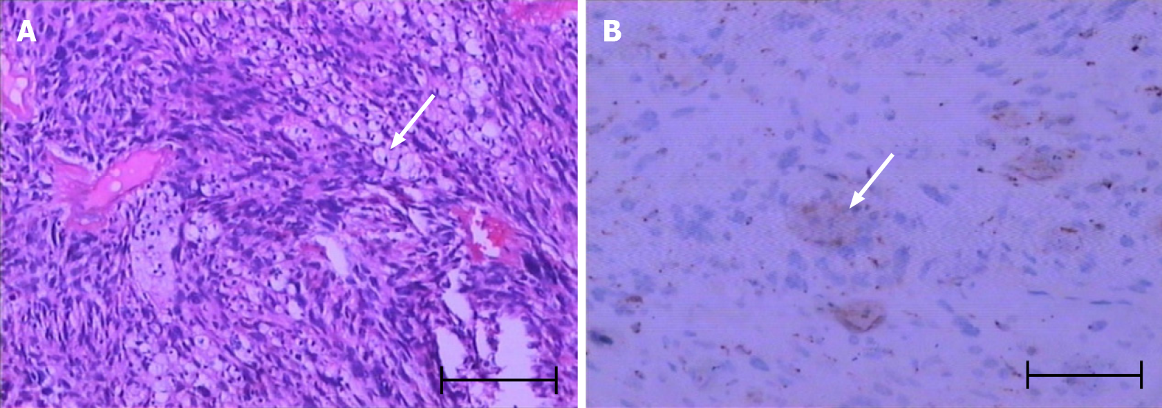
Figure 1 Pathological examination and immunohistochemical detection.
A: H&E staining showing obviously heterogeneous and fat spindle-shaped cells, arranged in intertwined and bundle-like patterns (arrow); B: Immunohistochemical staining establishing the spindle cell origin of the abnormal cell population: CD68+ (arrow). Scale bar: 100 μm.
- Citation: Zhu YQ, Zhao GC, Zheng CX, Yuan L, Yuan GB. Managing spindle cell sarcoma with surgery and high-intensity focused ultrasound: A case report. World J Clin Cases 2023; 11(27): 6551-6557
- URL: https://www.wjgnet.com/2307-8960/full/v11/i27/6551.htm
- DOI: https://dx.doi.org/10.12998/wjcc.v11.i27.6551


