Copyright
©The Author(s) 2023.
World J Clin Cases. Sep 26, 2023; 11(27): 6537-6542
Published online Sep 26, 2023. doi: 10.12998/wjcc.v11.i27.6537
Published online Sep 26, 2023. doi: 10.12998/wjcc.v11.i27.6537
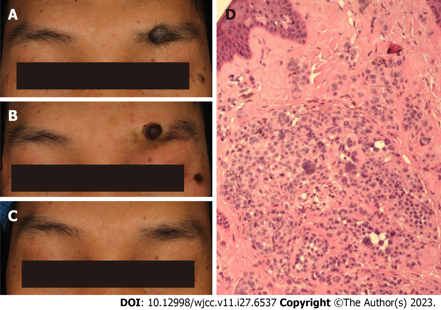
Figure 1 35-year-old male with an eyebrow intradermal nevus.
A: Before treatment, the lesion, which was located on the left brow arch, was a 13 mm round nodule with hair growth and a grey-brown appearance; B: Immediately after the surgery, there was a depressed wound on the left eyebrow arch with a dark brown scab attached, measuring approximately 10 mm; C: Thirteen months after the surgery, a light red macule measuring approximately 3 mm was observed at the site of the original skin lesion. There were no signs of depression or hypertrophic scarring, and the eyebrow was still intact; D: Under the microscope, the nevus cells in the dermis were arranged in a solid mass structure and were round, polygonal, spindle-shaped, or dendritic. The nuclei were vesicular and contained a few melanin granules.
- Citation: Liu C, Liang JL, Yu JL, Hu Q, Li CX. Successful treatment of eyebrow intradermal nevi by shearing combined with electrocautery and curettage: Two case reports. World J Clin Cases 2023; 11(27): 6537-6542
- URL: https://www.wjgnet.com/2307-8960/full/v11/i27/6537.htm
- DOI: https://dx.doi.org/10.12998/wjcc.v11.i27.6537


