Copyright
©The Author(s) 2023.
World J Clin Cases. Sep 26, 2023; 11(27): 6523-6530
Published online Sep 26, 2023. doi: 10.12998/wjcc.v11.i27.6523
Published online Sep 26, 2023. doi: 10.12998/wjcc.v11.i27.6523
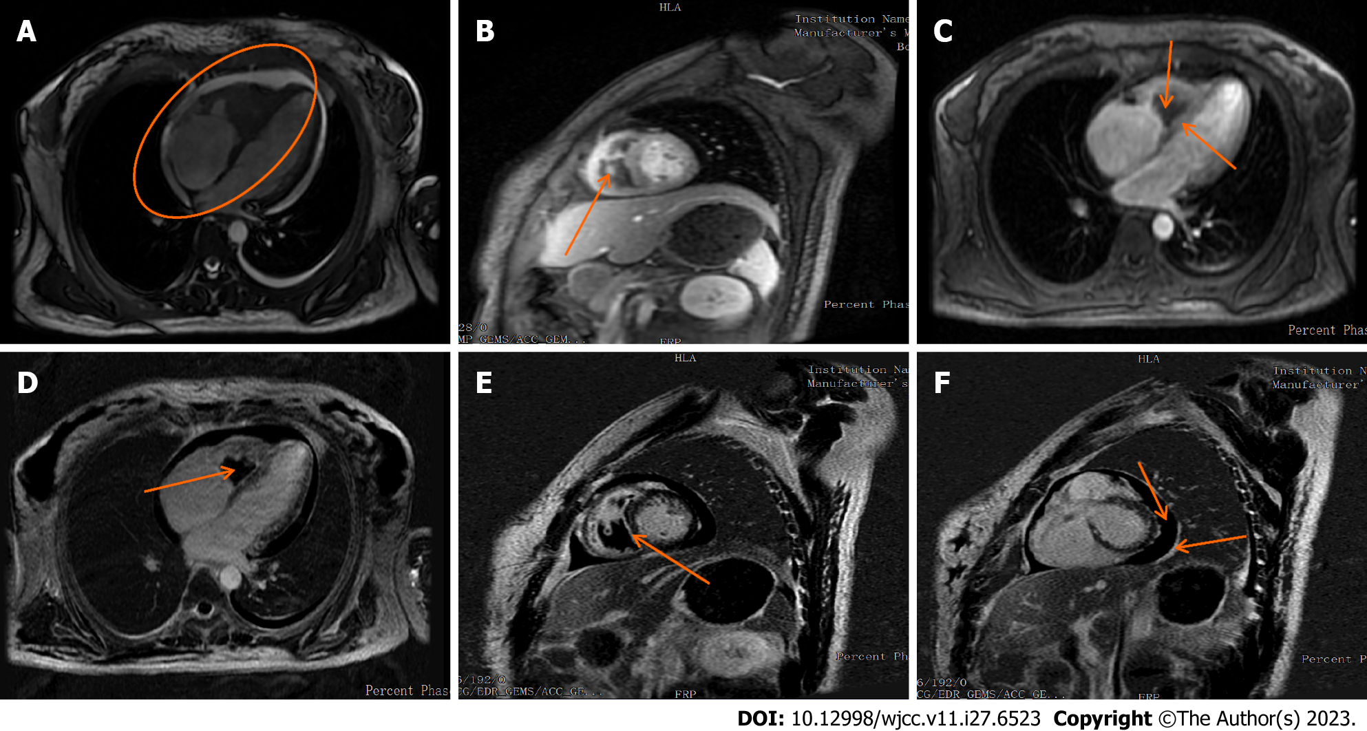
Figure 4 Cardiovascular magnetic resonance imaging illustrating cardiac changes.
A: Enlarged right atrial volume and thickened right ventricular endocardium; B: Central right ventricle displaying a low-signal filling defect; C: Myocardial first-perfusion highlighting a perfusion defect in the right ventricle with arcuate hypoperfusion areas located in the septum and right ventricular subendocardium; D: Delayed enhancement image illustrating no right ventricular enhancement; E: Delayed enhancement depicting high signal shadows present in the ventricular septum and on both ventricular walls; F: Delayed enhancement image showing minimal pericardial effusion and thickened pericardium.
- Citation: He JL, Liu XY, Zhang Y, Niu L, Li XL, Xie XY, Kang YT, Yang LQ, Cai ZY, Long H, Ye GF, Zou JX. Eosinophilic granulomatosis with polyangiitis, asthma as the first symptom, and subsequent Loeffler endocarditis: A case report. World J Clin Cases 2023; 11(27): 6523-6530
- URL: https://www.wjgnet.com/2307-8960/full/v11/i27/6523.htm
- DOI: https://dx.doi.org/10.12998/wjcc.v11.i27.6523


