Copyright
©The Author(s) 2023.
World J Clin Cases. Sep 26, 2023; 11(27): 6523-6530
Published online Sep 26, 2023. doi: 10.12998/wjcc.v11.i27.6523
Published online Sep 26, 2023. doi: 10.12998/wjcc.v11.i27.6523
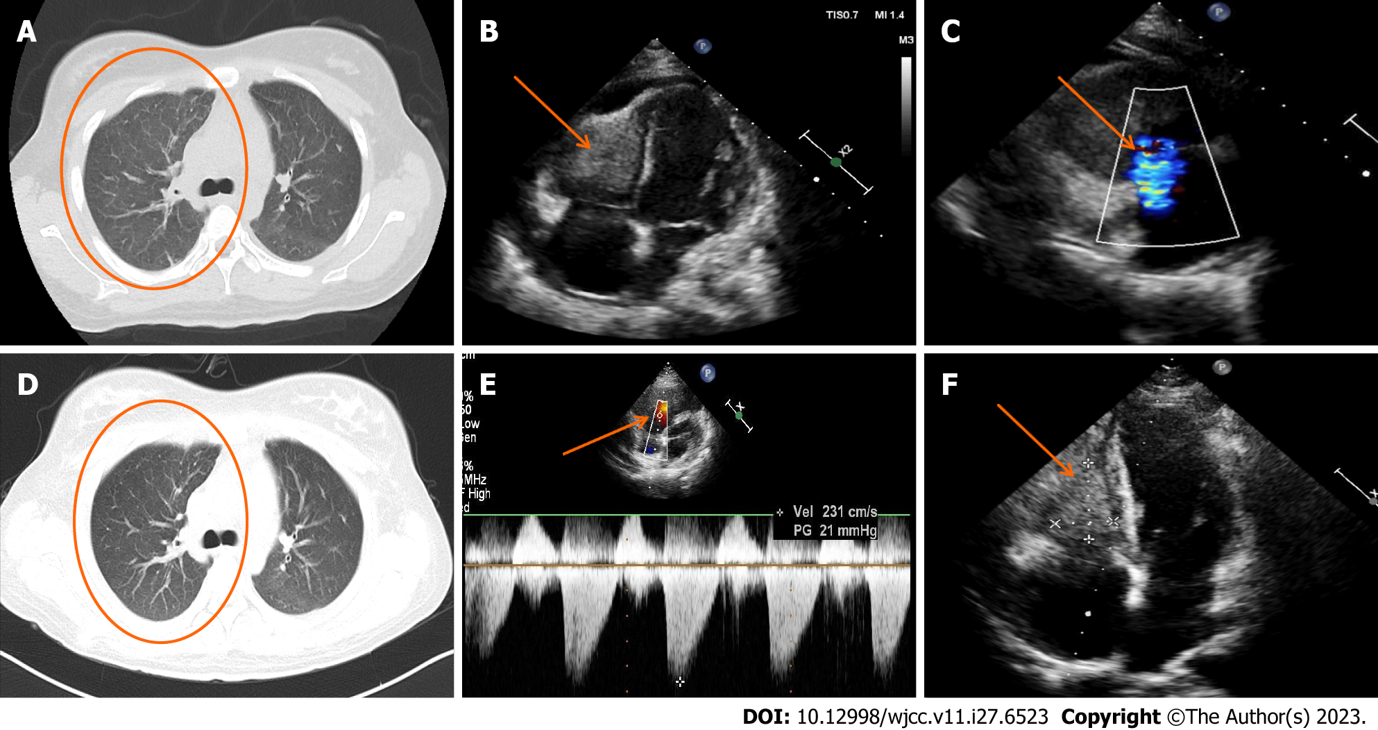
Figure 3 Comparative echocardiogram and chest computed tomography imaging pre- and post-treatment.
A: Chest computed tomography (CT; lung window image) from March 30, 2023; B and C: Pre-treatment echocardiogram illustrating right ventricular enlargement and a hypoechoic mass (dimensions: 25 mm × 24 mm × 38 mm) in the right ventricular wall; C: Significant regurgitation observed at the tricuspid valve; D: Comparative chest CT from April 7, 2023, post one week of glucocorticoid treatment, displaying diminished exudative foci in both lungs; E: Post-treatment echocardiogram indicating a tricuspid regurgitation peak flow rate of 231 cm/s, and a differential pressure of 21 mmHg, suggestive of regular systolic right ventricular pressure; F: Reduced echo size of right ventricular thrombus post-treatment.
- Citation: He JL, Liu XY, Zhang Y, Niu L, Li XL, Xie XY, Kang YT, Yang LQ, Cai ZY, Long H, Ye GF, Zou JX. Eosinophilic granulomatosis with polyangiitis, asthma as the first symptom, and subsequent Loeffler endocarditis: A case report. World J Clin Cases 2023; 11(27): 6523-6530
- URL: https://www.wjgnet.com/2307-8960/full/v11/i27/6523.htm
- DOI: https://dx.doi.org/10.12998/wjcc.v11.i27.6523


