Copyright
©The Author(s) 2023.
World J Clin Cases. Sep 26, 2023; 11(27): 6363-6373
Published online Sep 26, 2023. doi: 10.12998/wjcc.v11.i27.6363
Published online Sep 26, 2023. doi: 10.12998/wjcc.v11.i27.6363
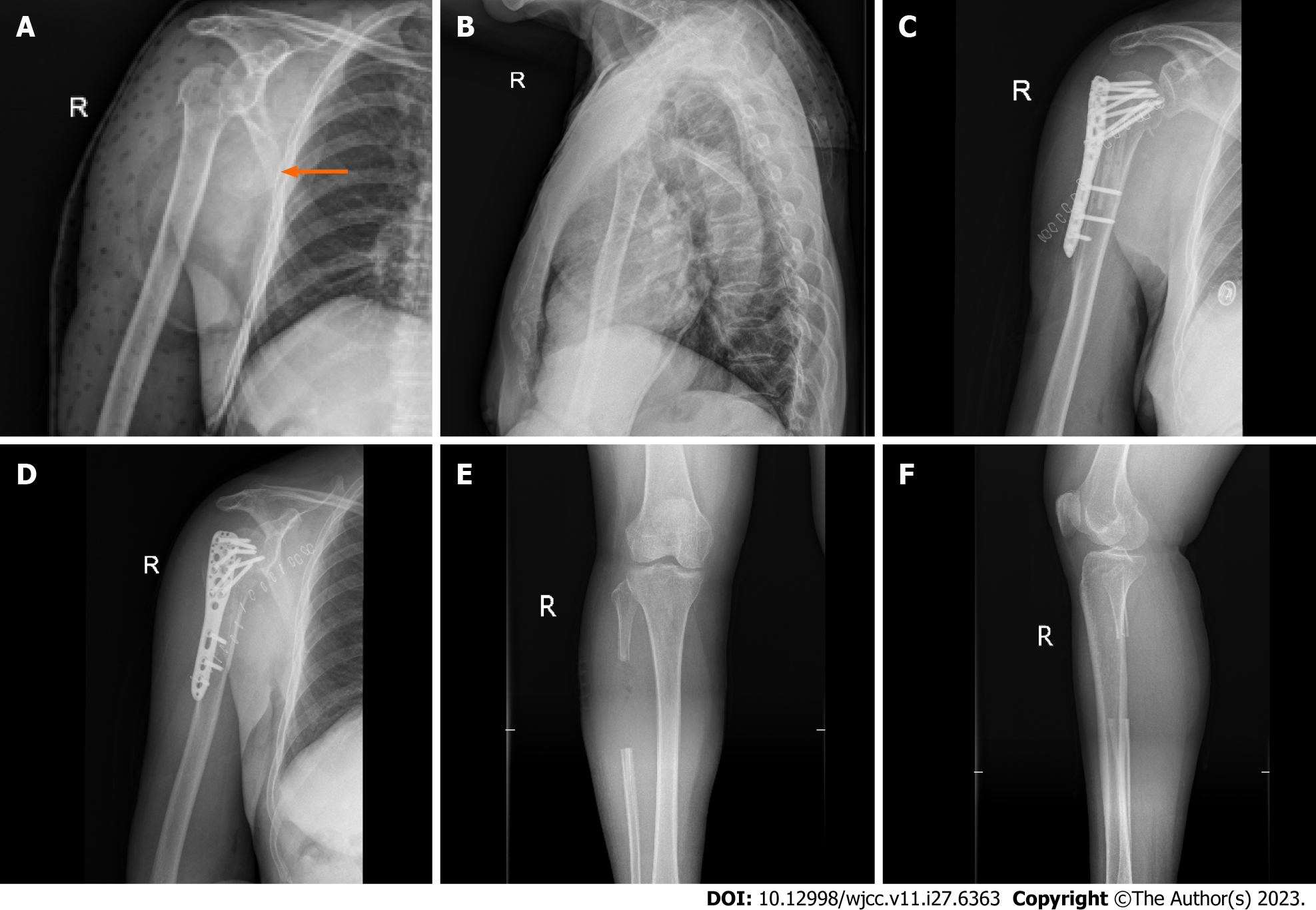
Figure 2 Radiographs of a typical case.
A and B: Anteroposterior and lateral radiographs of the proximal humerus before surgery (orange arrow indicates the displaced humeral head); C and D: Anteroposterior and lateral radiographs of the proximal humerus after surgery; E and F: Anteroposterior radiographs of the tibia and fibula after removal of the fibular segment.
- Citation: Liu N, Wang BG, Zhang LF. Treatment of proximal humeral fractures accompanied by medial calcar fractures using fibular autografts: A retrospective, comparative cohort study. World J Clin Cases 2023; 11(27): 6363-6373
- URL: https://www.wjgnet.com/2307-8960/full/v11/i27/6363.htm
- DOI: https://dx.doi.org/10.12998/wjcc.v11.i27.6363


