Copyright
©The Author(s) 2023.
World J Clin Cases. Sep 16, 2023; 11(26): 6289-6297
Published online Sep 16, 2023. doi: 10.12998/wjcc.v11.i26.6289
Published online Sep 16, 2023. doi: 10.12998/wjcc.v11.i26.6289
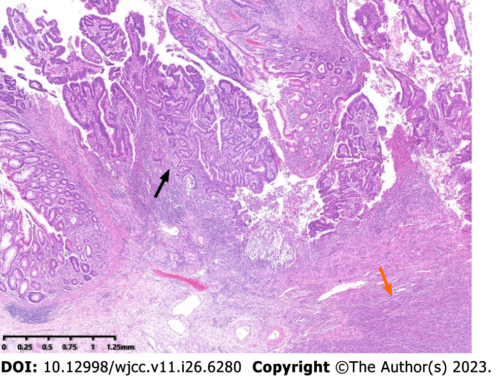
Figure 2 Low power view of the mass.
Microscopic examination disclosed that the tumor was composed of two components, adjacent to each other but relatively independent and showing infiltrative glands (black arrow) with underlying lymphoid proliferation (orange arrow) (Hematoxylin and eosin, original magnificent 20×).
- Citation: Jiang M, Yuan XP. Collision tumor of primary malignant lymphoma and adenocarcinoma in the colon diagnosed by molecular pathology: A case report and literature review. World J Clin Cases 2023; 11(26): 6289-6297
- URL: https://www.wjgnet.com/2307-8960/full/v11/i26/6289.htm
- DOI: https://dx.doi.org/10.12998/wjcc.v11.i26.6289


