Copyright
©The Author(s) 2023.
World J Clin Cases. Sep 16, 2023; 11(26): 6274-6279
Published online Sep 16, 2023. doi: 10.12998/wjcc.v11.i26.6274
Published online Sep 16, 2023. doi: 10.12998/wjcc.v11.i26.6274
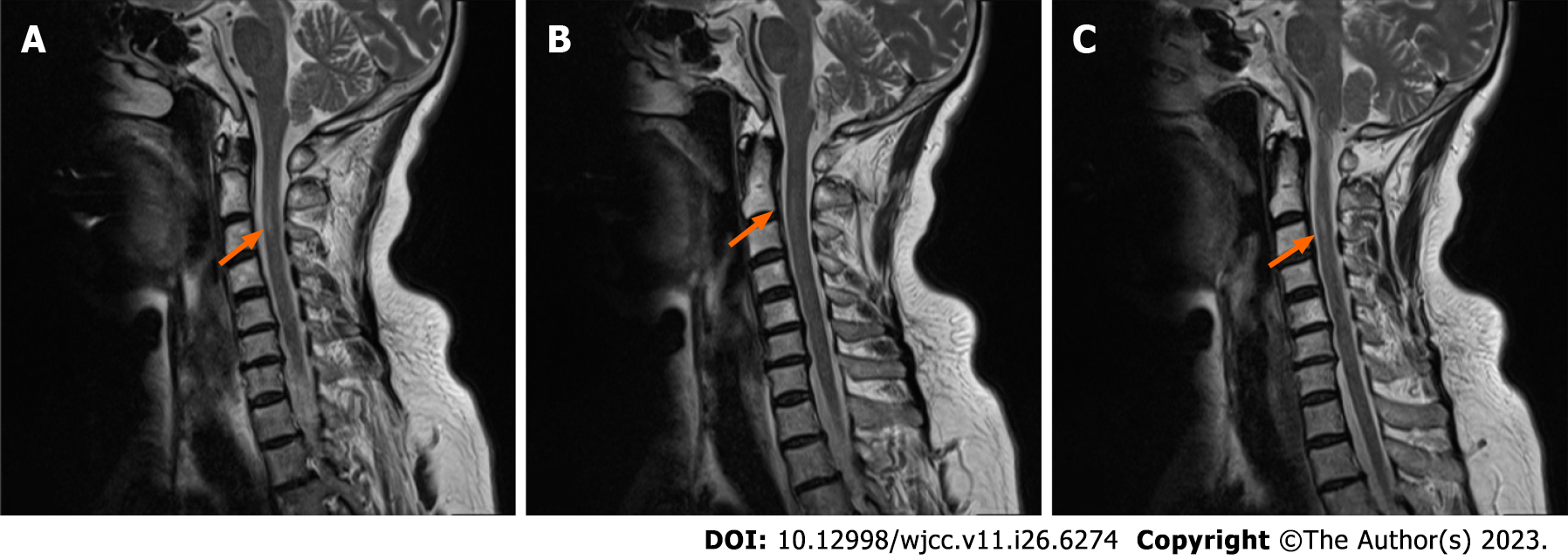
Figure 2 Magnetic resonance imaging of patient's cervical spine.
A-C: Sagittal plane magnetic resonance imaging shows the lesion site (orange arrows) and T2WI shows multiple discs with a reduced signal and a slightly longer lamellar T2 signal in the medulla at the level of the C2–C3 vertebral body. Straightening of the normal curvature of the cervical spine in images at different sagittal positions.
- Citation: Liu H, Lv HG, Zhang R. Variant of Guillain-Barré syndrome with anti-sulfatide antibody positivity and spinal cord involvement: A case report. World J Clin Cases 2023; 11(26): 6274-6279
- URL: https://www.wjgnet.com/2307-8960/full/v11/i26/6274.htm
- DOI: https://dx.doi.org/10.12998/wjcc.v11.i26.6274


