Copyright
©The Author(s) 2023.
World J Clin Cases. Sep 16, 2023; 11(26): 6268-6273
Published online Sep 16, 2023. doi: 10.12998/wjcc.v11.i26.6268
Published online Sep 16, 2023. doi: 10.12998/wjcc.v11.i26.6268
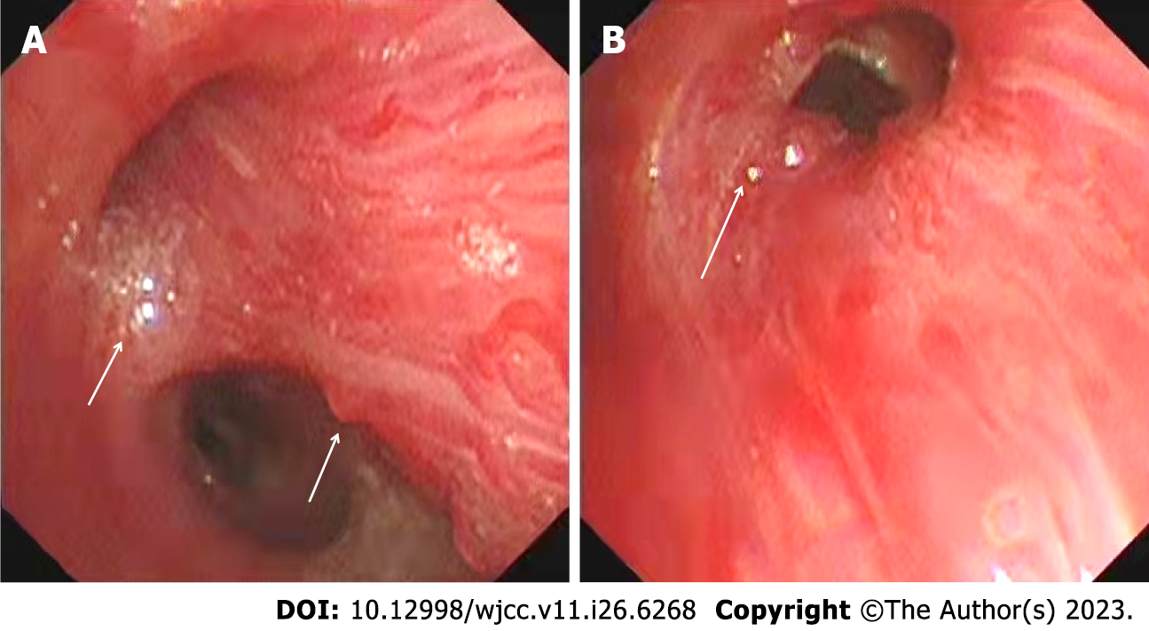
Figure 1 Fiberoptic bronchoscopy.
A: Multiple nodular protrusions and scar formation in the right middle segment of the bronchial mucosa (arrow); B: Nodular protuberances in the basal segment of the right lower lobe (arrow), where the mucosa could easily bleed.
- Citation: Wang F, Tang J, Peng M, Huang PJ, Zhao LJ, Zhang YY, Wang T. Recurrent hemoptysis in pediatric bronchial Dieulafoy’s disease with inferior phrenic artery supply: A case report. World J Clin Cases 2023; 11(26): 6268-6273
- URL: https://www.wjgnet.com/2307-8960/full/v11/i26/6268.htm
- DOI: https://dx.doi.org/10.12998/wjcc.v11.i26.6268


