Copyright
©The Author(s) 2023.
World J Clin Cases. Sep 16, 2023; 11(26): 6252-6261
Published online Sep 16, 2023. doi: 10.12998/wjcc.v11.i26.6252
Published online Sep 16, 2023. doi: 10.12998/wjcc.v11.i26.6252
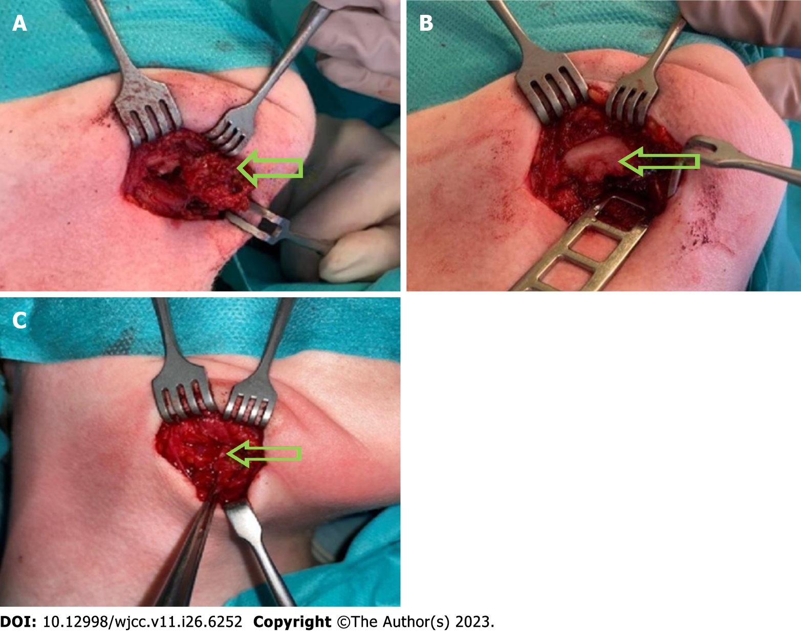
Figure 5 Intraoperative picture.
A: Intraoperative picture of submandibular, angiomatoid fibrous histiocytoma, tumor is marked with an arrow; B: Intraoperative picture of a destroyed external layer of the mandible, it is marked with an arrow; C: Intraoperative view of the second procedure, no AFH irregularities were found. Tissue scar are marked with arrows.
- Citation: Michcik A, Bień M, Wojciechowska B, Polcyn A, Garbacewicz Ł, Kowalski J, Drogoszewska B. Difficulties in diagnosing angiomatoid fibrous histiocytoma of the head and neck region: A case report. World J Clin Cases 2023; 11(26): 6252-6261
- URL: https://www.wjgnet.com/2307-8960/full/v11/i26/6252.htm
- DOI: https://dx.doi.org/10.12998/wjcc.v11.i26.6252


