Copyright
©The Author(s) 2023.
World J Clin Cases. Sep 16, 2023; 11(26): 6252-6261
Published online Sep 16, 2023. doi: 10.12998/wjcc.v11.i26.6252
Published online Sep 16, 2023. doi: 10.12998/wjcc.v11.i26.6252
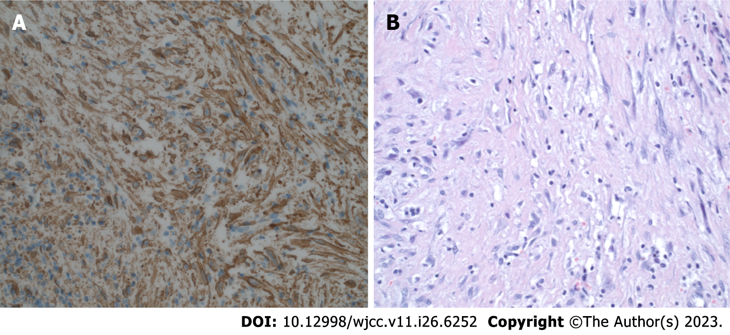
Figure 4 Histopathological diagnosis.
A: Diffuse expression of smooth muscle actin. Excision, magnification 400 ×; B: Excision (400 ×, hematoxylin and eosin staining). Short spindled myoid and ovoid, histiocyte-like cells within a somewhat myxoid background.
- Citation: Michcik A, Bień M, Wojciechowska B, Polcyn A, Garbacewicz Ł, Kowalski J, Drogoszewska B. Difficulties in diagnosing angiomatoid fibrous histiocytoma of the head and neck region: A case report. World J Clin Cases 2023; 11(26): 6252-6261
- URL: https://www.wjgnet.com/2307-8960/full/v11/i26/6252.htm
- DOI: https://dx.doi.org/10.12998/wjcc.v11.i26.6252


