Copyright
©The Author(s) 2023.
World J Clin Cases. Sep 16, 2023; 11(26): 6252-6261
Published online Sep 16, 2023. doi: 10.12998/wjcc.v11.i26.6252
Published online Sep 16, 2023. doi: 10.12998/wjcc.v11.i26.6252
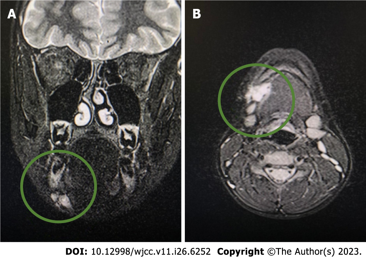
Figure 1 Computed tomography.
A: Focal change on the right side of the submandibular space initially diagnosed as a venous malformation; B: Visible irregular outline of a moderately hyperintense area of 18 mm × 25 mm × 13 mm.
- Citation: Michcik A, Bień M, Wojciechowska B, Polcyn A, Garbacewicz Ł, Kowalski J, Drogoszewska B. Difficulties in diagnosing angiomatoid fibrous histiocytoma of the head and neck region: A case report. World J Clin Cases 2023; 11(26): 6252-6261
- URL: https://www.wjgnet.com/2307-8960/full/v11/i26/6252.htm
- DOI: https://dx.doi.org/10.12998/wjcc.v11.i26.6252


