Copyright
©The Author(s) 2023.
World J Clin Cases. Sep 16, 2023; 11(26): 6240-6245
Published online Sep 16, 2023. doi: 10.12998/wjcc.v11.i26.6240
Published online Sep 16, 2023. doi: 10.12998/wjcc.v11.i26.6240
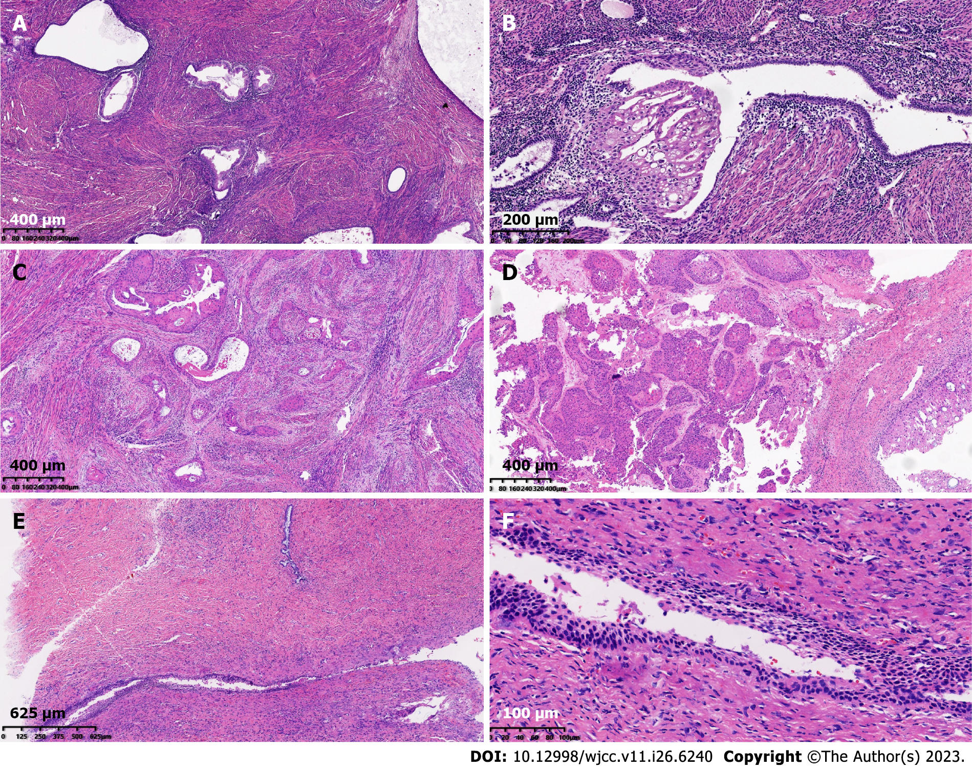
Figure 2 Morphological features.
A: Endometrial glands appeared in the myometrium [Hematoxylin and eosin (HE) 10 ×]; B: Some ectopic endometrial glands showed metaplasia in squamous epithelium (HE 10 ×); C: Tumor cells showed differentiation characteristics in squamous epithelium, arrayed as nests and infiltrated with a large amount of keratinization (HE 10 ×); D: The cystic wall of a solid cystic mass in the right ovary (HE 10 ×); E and F: Normal squamous epithelial and cervical glands were observed in cervical tissues (HE 4 ×, 10 ×).
- Citation: Cai Z, Yang GL, Li Q, Zeng L, Li LX, Song YP, Liu FR. Squamous cell carcinoma associated with endometriosis in the uterus and ovaries: A case report. World J Clin Cases 2023; 11(26): 6240-6245
- URL: https://www.wjgnet.com/2307-8960/full/v11/i26/6240.htm
- DOI: https://dx.doi.org/10.12998/wjcc.v11.i26.6240


