Copyright
©The Author(s) 2023.
World J Clin Cases. Sep 16, 2023; 11(26): 6223-6230
Published online Sep 16, 2023. doi: 10.12998/wjcc.v11.i26.6223
Published online Sep 16, 2023. doi: 10.12998/wjcc.v11.i26.6223
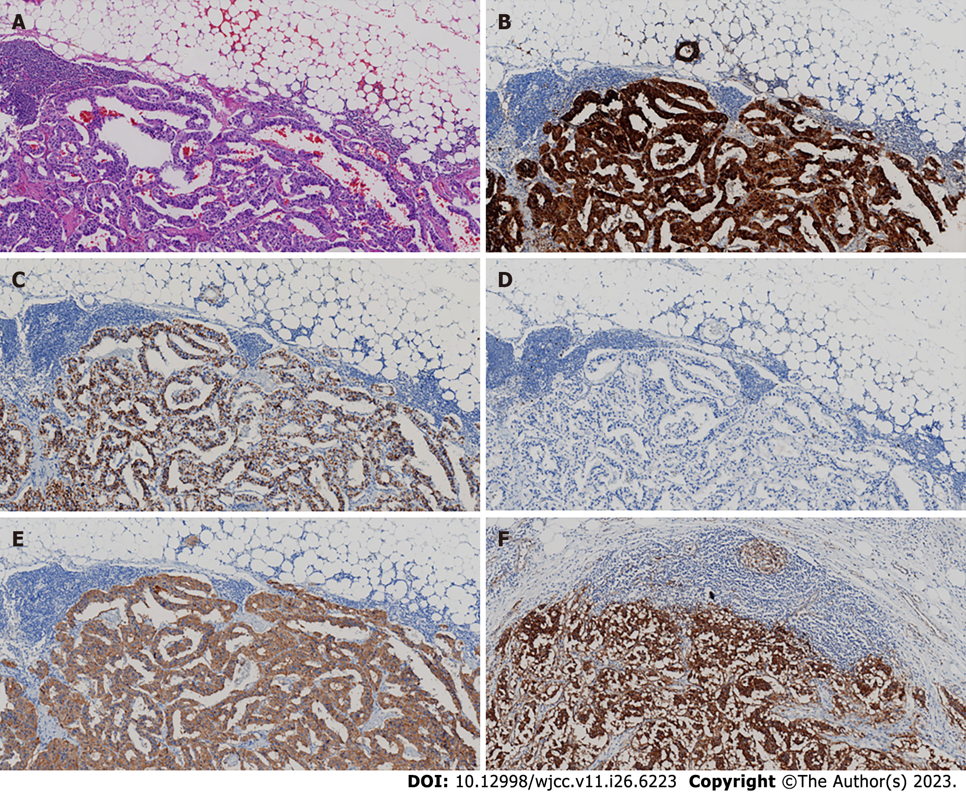
Figure 3 Pathological smears of tumor tissue after various immunohistochemical staining.
A: Hematoxylin and eosin (× 100); B: Chromogranin A (× 100); C: Gastrin (× 100); D: Ki-67 (× 100); E: Syn (× 100); F: Somatostatin receptors 2 (× 100). B and C suggest a gastrinoma; D represents proliferative activity; E and F suggests a neuroendocrine tumor.
- Citation: Zhang JM, Zheng CW, Li XW, Fang ZY, Yu MX, Shen HY, Ji X. Typical Zollinger-Ellison syndrome-atypical location of gastrinoma and absence of hypergastrinemia: A case report and review of literature. World J Clin Cases 2023; 11(26): 6223-6230
- URL: https://www.wjgnet.com/2307-8960/full/v11/i26/6223.htm
- DOI: https://dx.doi.org/10.12998/wjcc.v11.i26.6223


