Copyright
©The Author(s) 2023.
World J Clin Cases. Sep 16, 2023; 11(26): 6159-6164
Published online Sep 16, 2023. doi: 10.12998/wjcc.v11.i26.6159
Published online Sep 16, 2023. doi: 10.12998/wjcc.v11.i26.6159
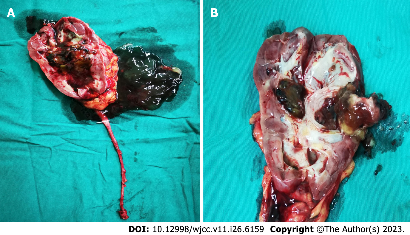
Figure 3 Gross pathology of the resected right kidney and ureter.
A: A large amount of gelatinous mucinous fluid was found in the renal pelvis and calyces; B: A hard solid mass about 2.5 cm 2.0 cm in size was found in the middle pole of the kidney. Furthermore, there was a stone about 1 cm in the mass and a papillary neoplasm below the mass.
- Citation: Li LL, Song PX, Xing DF, Liu K. Early diagnosis of renal pelvis villous adenoma: A case report. World J Clin Cases 2023; 11(26): 6159-6164
- URL: https://www.wjgnet.com/2307-8960/full/v11/i26/6159.htm
- DOI: https://dx.doi.org/10.12998/wjcc.v11.i26.6159


