Copyright
©The Author(s) 2023.
World J Clin Cases. Sep 16, 2023; 11(26): 6147-6153
Published online Sep 16, 2023. doi: 10.12998/wjcc.v11.i26.6147
Published online Sep 16, 2023. doi: 10.12998/wjcc.v11.i26.6147
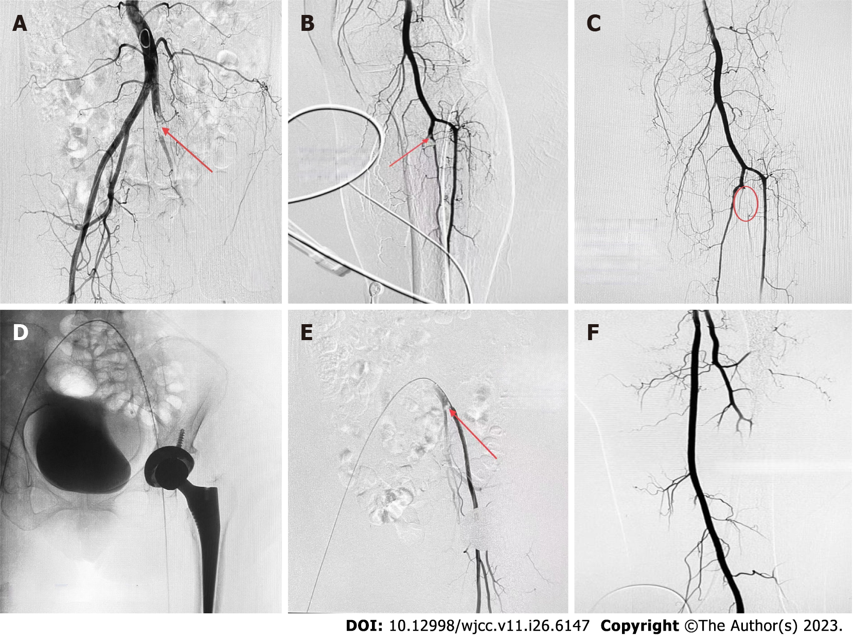
Figure 2 Imaging evidence collected during the percutaneous arterial angiograph.
A: Thrombosis in the distal common iliac artery; B: Thromboses in the tibiofibular trunk; C: Lower limb arteriography imaging after thrombectomy and thrombolysis; D Insertion of thrombolysis catheter into common iliac artery; E The iliac artery thromboses became smaller after the repeat angiography; F Angiography of left common iliac artery and internal/external iliac artery after femoral artery thrombectomy.
- Citation: Lv FF, Li MY, Qu W, Jiang ZS. Rivaroxaban for the treatment of heparin-induced thrombocytopenia with thrombosis in a patient undergoing artificial hip arthroplasty: A case report. World J Clin Cases 2023; 11(26): 6147-6153
- URL: https://www.wjgnet.com/2307-8960/full/v11/i26/6147.htm
- DOI: https://dx.doi.org/10.12998/wjcc.v11.i26.6147


