Copyright
©The Author(s) 2023.
World J Clin Cases. Sep 16, 2023; 11(26): 6091-6104
Published online Sep 16, 2023. doi: 10.12998/wjcc.v11.i26.6091
Published online Sep 16, 2023. doi: 10.12998/wjcc.v11.i26.6091
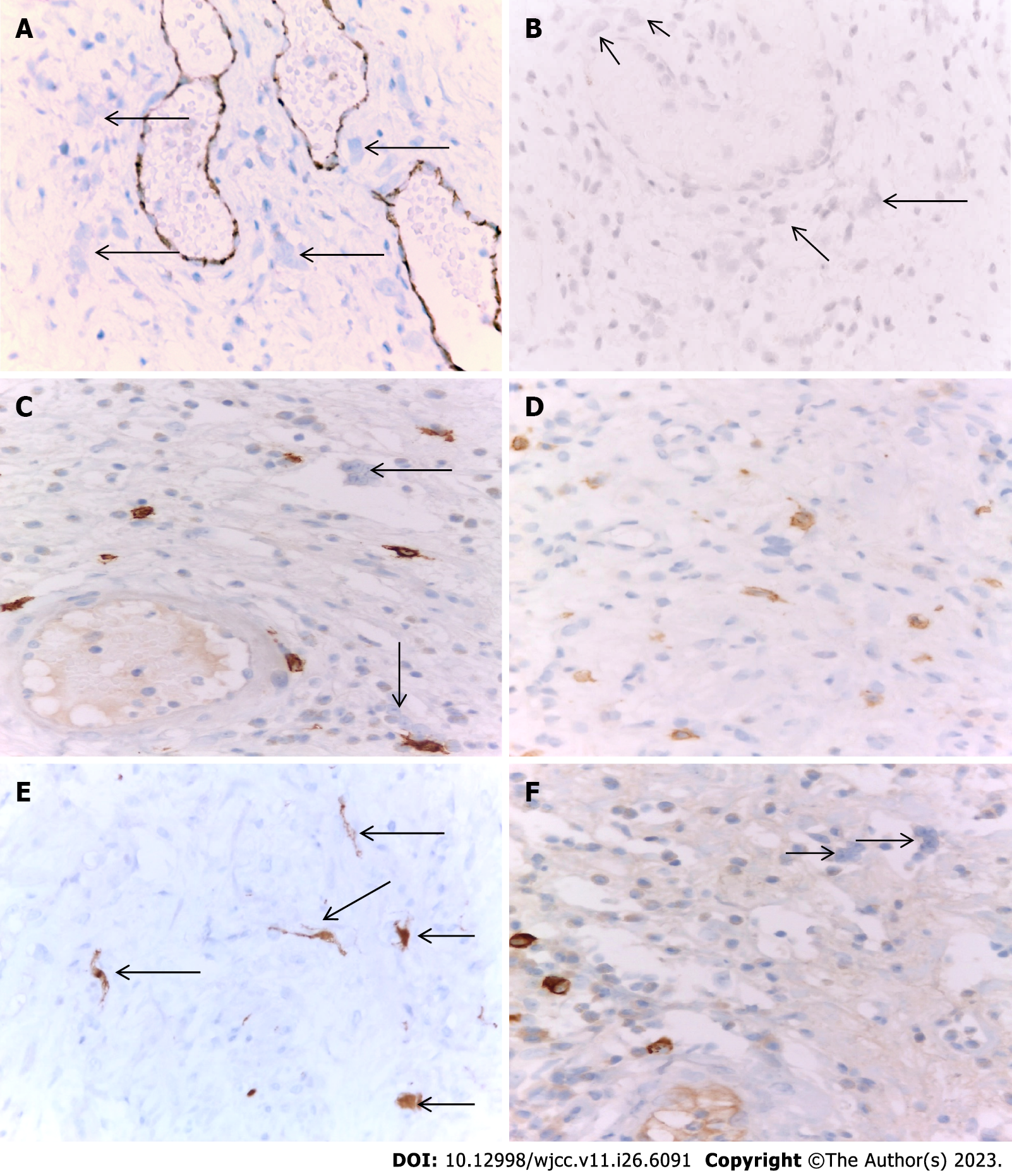
Figure 5 Immunohistochemical examination of urothelial carcinoma of the bladder.
A: Presence of several pericapillary multinucleated giant cells (MGCs) in the mucosa, which were CD31-negative (arrow); the endothelial cells of the surrounding capillaries served for internal positive control. immunohistochemical (IHC), CD31 × 400; B: Presence of several pericapillary MGCs in the mucosa, which were CD10-negative (arrow). IHC, CD10 × 400; C: The presence of two pericapillary MGCs in the bladder’s mucosa, which were found negative for C-KIT (arrow); positively labeled pericapillary mast cells were used for internal positive control. IHC, C-KIT × 630; D: MGCs in the bladder mucosa were always accompanied by many pericapillary C-KIT positive mast cells. IHC, C-KIT × 630; E: Giant cells were p16-positive (arrow). IHC, p16 × 400; F: Presence of two pericapillary MGCs in the mucosa that are negative immunoglobulin G4 (IG G4) for IG G4; several positively labeled pericapillary plasma cells were used for internal positive control. IHC, IG G4 × 630.
- Citation: Gulinac M, Velikova T, Dikov D. Multinucleated giant cells of bladder mucosa are modified telocytes: Diagnostic and immunohistochemistry algorithm and relation to PD-L1 expression score. World J Clin Cases 2023; 11(26): 6091-6104
- URL: https://www.wjgnet.com/2307-8960/full/v11/i26/6091.htm
- DOI: https://dx.doi.org/10.12998/wjcc.v11.i26.6091


