Copyright
©The Author(s) 2023.
World J Clin Cases. Sep 16, 2023; 11(26): 6091-6104
Published online Sep 16, 2023. doi: 10.12998/wjcc.v11.i26.6091
Published online Sep 16, 2023. doi: 10.12998/wjcc.v11.i26.6091
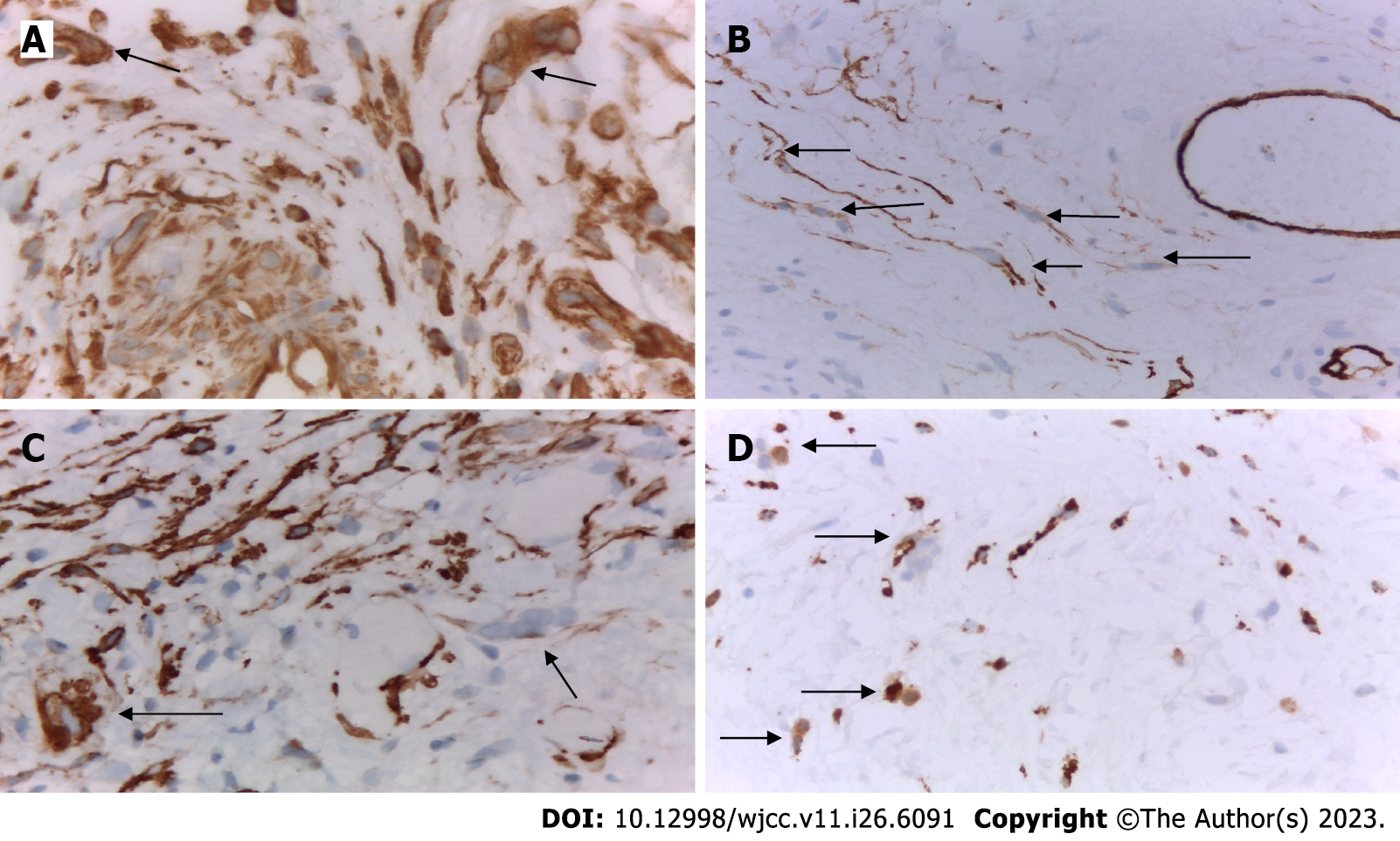
Figure 4 Immunohistochemical examination of urothelial carcinoma of the bladder.
A: Presence of two pericapillary multinucleated giant cells (MGCs) in the mucosa that were vimentin-positive (arrow). Immunohistochemical (IHC), VIMENTIN × 630; B: Presence of several pericapillary MGCs in the mucosa, which were CD34 + positive (arrow); the cells in the wall of the surrounding capillaries served for internal positive control. IHC, CD34 × 400; C: Presence of two pericapillary MGCs in the mucosa, the growths of which were positive for smooth muscle actin (arrow); the cells in the wall of the surrounding capillaries served as an internal positive control. IHC, SMA × 400; D: Presence of several MGCs in the mucosa whose cytoplasm was positive for the macrophage marker CD68 (arrow); mononuclear macrophages the context of the inflammatory infiltrate served as an internal positive control. IHC, CD68 × 400.
- Citation: Gulinac M, Velikova T, Dikov D. Multinucleated giant cells of bladder mucosa are modified telocytes: Diagnostic and immunohistochemistry algorithm and relation to PD-L1 expression score. World J Clin Cases 2023; 11(26): 6091-6104
- URL: https://www.wjgnet.com/2307-8960/full/v11/i26/6091.htm
- DOI: https://dx.doi.org/10.12998/wjcc.v11.i26.6091


