Copyright
©The Author(s) 2023.
World J Clin Cases. Sep 16, 2023; 11(26): 6091-6104
Published online Sep 16, 2023. doi: 10.12998/wjcc.v11.i26.6091
Published online Sep 16, 2023. doi: 10.12998/wjcc.v11.i26.6091
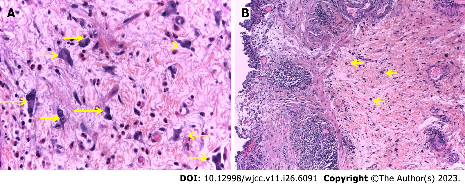
Figure 3 Conventional histological examination of urothelial carcinoma of the bladder.
A: Presence of atypical mono-, bi- and multin urothelial carcinoma (UC) leated giant fibroblast-like stromal cells (arrow) in the vesicular lamina propria adjacent to non-invasive papillary low-grade (LG) UC. A nonspecific stromal inflammatory response rich in plasma cells and eosinophilic leukocytes accompany them. hematoxylin-eosin, 400 ×; B: Presence of multinucleated giant cells in the bladder lamina propria (arrow) in close proximity to tertiary lymphoid structures of type 2 and 3 (2nd and 3rd degree) and with non-invasive papillary LG UC. hematoxylin-eosin, 100 ×.
- Citation: Gulinac M, Velikova T, Dikov D. Multinucleated giant cells of bladder mucosa are modified telocytes: Diagnostic and immunohistochemistry algorithm and relation to PD-L1 expression score. World J Clin Cases 2023; 11(26): 6091-6104
- URL: https://www.wjgnet.com/2307-8960/full/v11/i26/6091.htm
- DOI: https://dx.doi.org/10.12998/wjcc.v11.i26.6091


