Copyright
©The Author(s) 2023.
World J Clin Cases. Sep 6, 2023; 11(25): 6005-6011
Published online Sep 6, 2023. doi: 10.12998/wjcc.v11.i25.6005
Published online Sep 6, 2023. doi: 10.12998/wjcc.v11.i25.6005
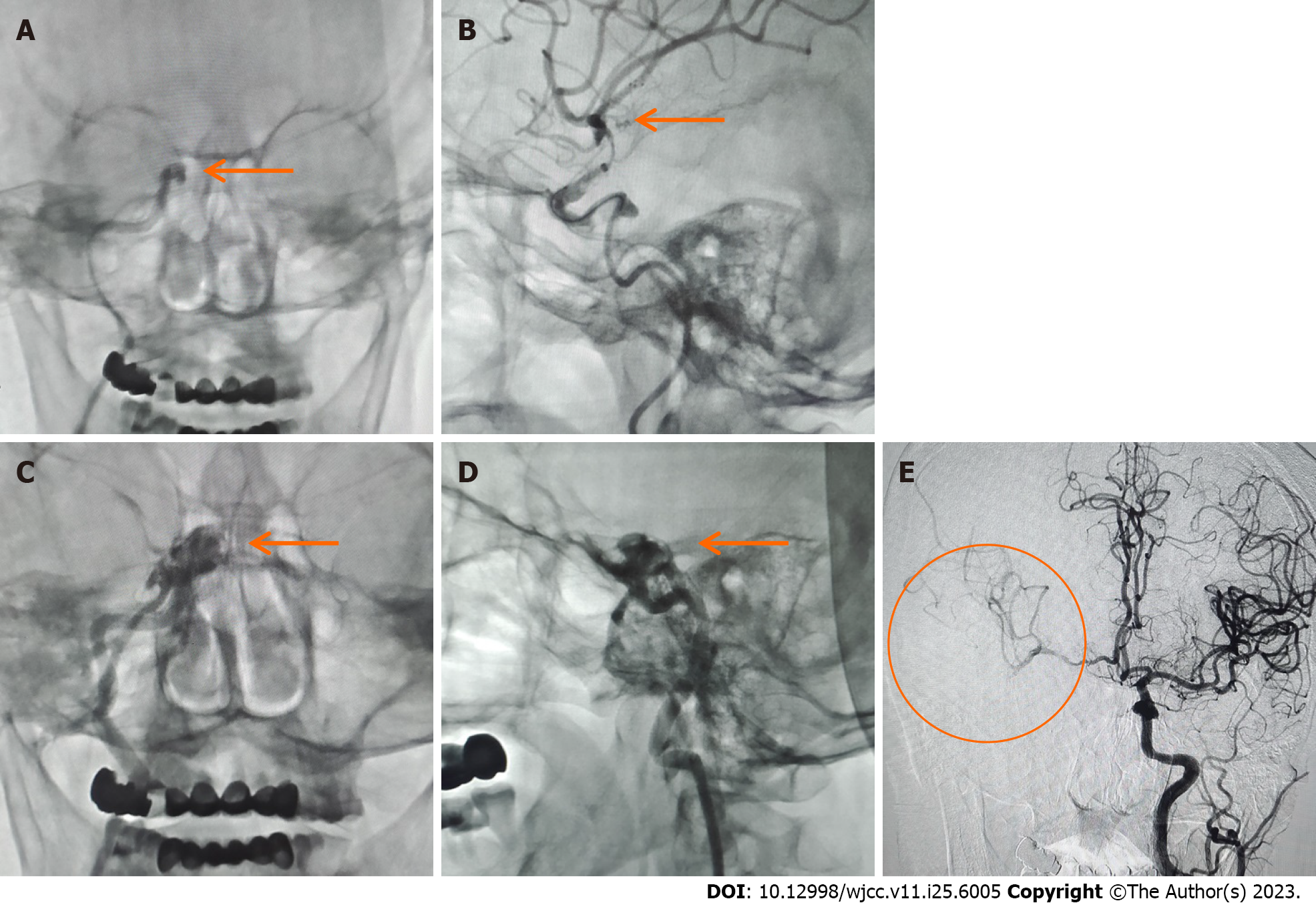
Figure 3 Thrombectomy and carotid-cavernous fistula formation.
A: After the aspiration thrombectomy, the digital subtraction angiography showed occlusion of the terminal bifurcation of the right internal carotid artery (arrow); B: After the first stent removal, angiography showed poor right middle cerebral artery (MCA) reflow and no carotid-cavernous fistula (CCF) (arrow); C and D: After a second stent thrombectomy, reexamination showed CCF (arrows); E: Reflow of the A1 segment of the right anterior cerebral artery to supply blood to the right MCA supply area (circle).
- Citation: Qu LZ, Dong GH, Zhu EB, Lin MQ, Liu GL, Guan HJ. Carotid-cavernous fistula following mechanical thrombectomy of the tortuous internal carotid artery: A case report. World J Clin Cases 2023; 11(25): 6005-6011
- URL: https://www.wjgnet.com/2307-8960/full/v11/i25/6005.htm
- DOI: https://dx.doi.org/10.12998/wjcc.v11.i25.6005


