Copyright
©The Author(s) 2023.
World J Clin Cases. Sep 6, 2023; 11(25): 5994-5999
Published online Sep 6, 2023. doi: 10.12998/wjcc.v11.i25.5994
Published online Sep 6, 2023. doi: 10.12998/wjcc.v11.i25.5994
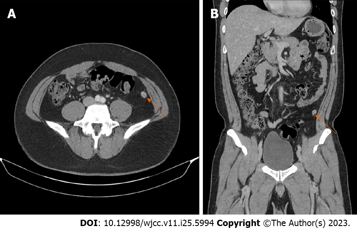
Figure 2 Abdominal computed tomography.
A: Axial view; B: Coronal view. Abdominal computed tomography at 6 mo postoperatively showed a 12 mm enhancing nodule at the left lower peritoneum (arrows).
- Citation: Chung JW, Kang JK, Lee EH, Chun SY, Ha YS, Lee JN, Kim TH, Kwon TG, Yoon GS. Single omental metastasis of renal cell carcinoma after radical nephrectomy: A case report. World J Clin Cases 2023; 11(25): 5994-5999
- URL: https://www.wjgnet.com/2307-8960/full/v11/i25/5994.htm
- DOI: https://dx.doi.org/10.12998/wjcc.v11.i25.5994


