Copyright
©The Author(s) 2023.
World J Clin Cases. Sep 6, 2023; 11(25): 5962-5969
Published online Sep 6, 2023. doi: 10.12998/wjcc.v11.i25.5962
Published online Sep 6, 2023. doi: 10.12998/wjcc.v11.i25.5962
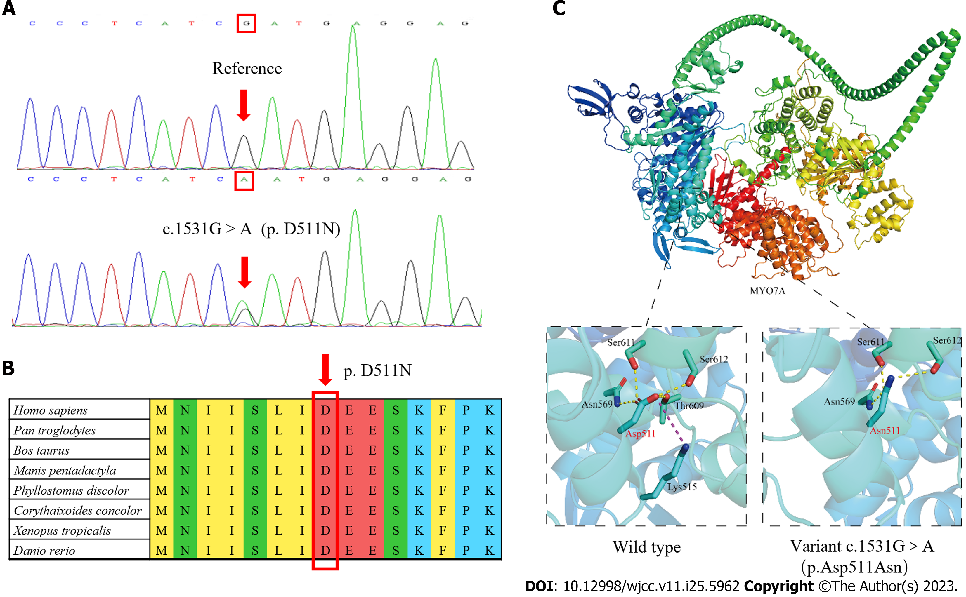
Figure 3 Location of nucleotide changes and functional analysis of the variant.
A: DNA sequence chromatograms. Arrows indicate the site of the mutation, which results in the p.D511N variant; B: Evolutionary conservation of Asp at position 511 (indicated by the arrow) on the MYO7A protein; C: The wild-type and variant (p.D511N) of the MYO7A protein. Yellow dashed lines represent hydrogen bonds between amino acids, and the purple dashed line represent electrostatic interaction between amino acids.
- Citation: Xia CF, Yan R, Su WW, Liu YH. Autosomal dominant non-syndromic hearing loss caused by a novel mutation in MYO7A: A case report and review of the literature. World J Clin Cases 2023; 11(25): 5962-5969
- URL: https://www.wjgnet.com/2307-8960/full/v11/i25/5962.htm
- DOI: https://dx.doi.org/10.12998/wjcc.v11.i25.5962


