Copyright
©The Author(s) 2023.
World J Clin Cases. Sep 6, 2023; 11(25): 5954-5961
Published online Sep 6, 2023. doi: 10.12998/wjcc.v11.i25.5954
Published online Sep 6, 2023. doi: 10.12998/wjcc.v11.i25.5954
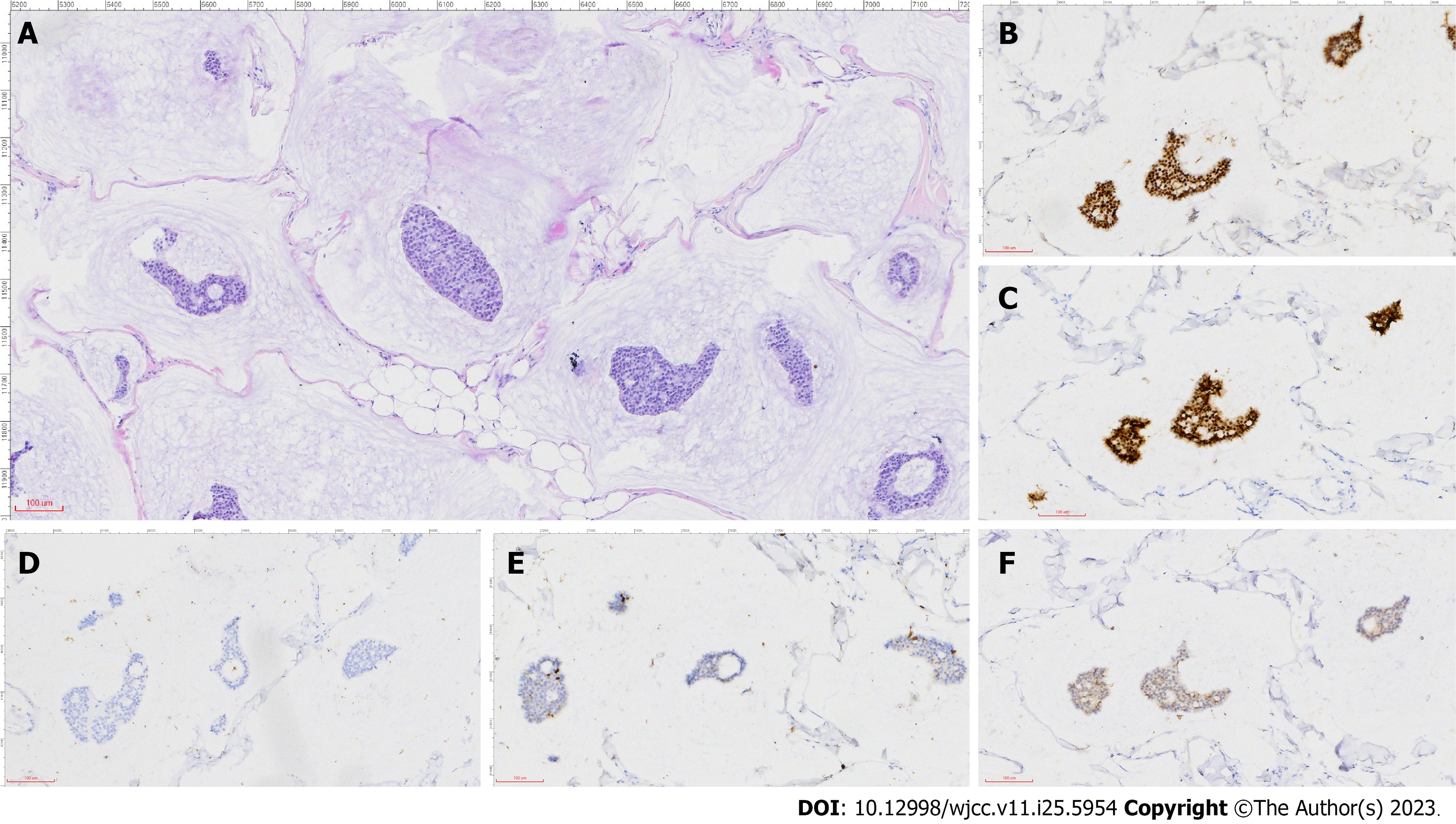
Figure 2 Pathology and immunohistochemistry analysis.
A: Image showing clusters of tumor cells suspended in abundant extracellular mucin and separated by ciliated fibrous intervals containing capillaries; B: Estrogen receptor (2+); C: Progesterone receptor (2+); D: AR (−); E: Ki-67 (5%); F: CerbB-2 (−) (scale: 100 μm; 200 ×).
- Citation: Sun Q, Liu XY, Zhang Q, Jiang H. Non-retroareolar male mucinous breast cancer without gynecomastia development in an elderly man: A case report. World J Clin Cases 2023; 11(25): 5954-5961
- URL: https://www.wjgnet.com/2307-8960/full/v11/i25/5954.htm
- DOI: https://dx.doi.org/10.12998/wjcc.v11.i25.5954


