Copyright
©The Author(s) 2023.
World J Clin Cases. Sep 6, 2023; 11(25): 5947-5953
Published online Sep 6, 2023. doi: 10.12998/wjcc.v11.i25.5947
Published online Sep 6, 2023. doi: 10.12998/wjcc.v11.i25.5947
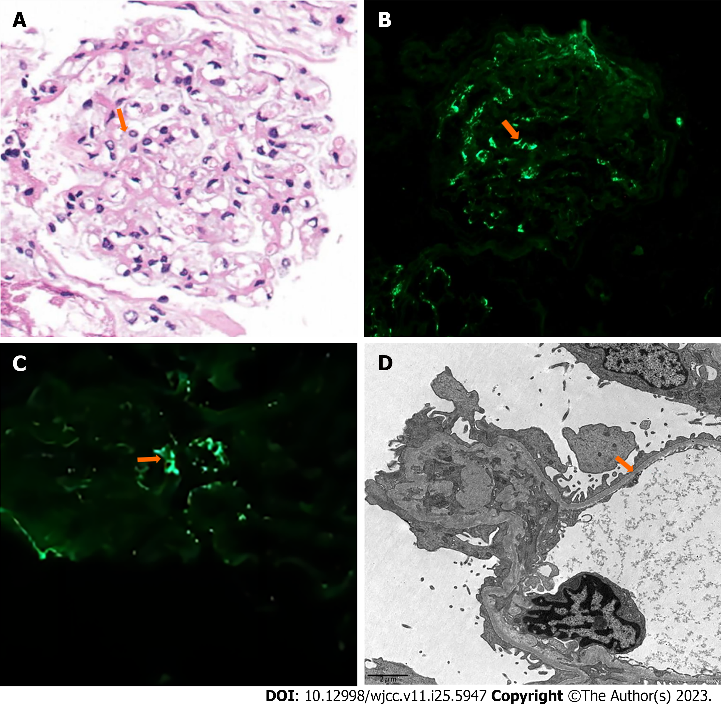
Figure 2 Findings of the proband’s renal biopsy.
A: Hematoxylin and eosin stain shows mild mesangial proliferation (as the arrow indicates). Original magnification 400 ×; B: Immunofluorescence shows deposition of immunoglobulin A (as the arrow indicates). Original magnification 400 ×; C: Immunofluorescence shows deposition of C3 (as the arrow indicates). Original magnification 400 ×; D: Electron microscopy shows diffuse thinning of the glomerular basement membrane (as the arrow indicates). Scale bars: 2 μm.
- Citation: Chen YT, Jiang WZ, Lu KD. Novel COL4A3 synonymous mutation causes Alport syndrome coexistent with immunoglobulin A nephropathy in a woman: A case report. World J Clin Cases 2023; 11(25): 5947-5953
- URL: https://www.wjgnet.com/2307-8960/full/v11/i25/5947.htm
- DOI: https://dx.doi.org/10.12998/wjcc.v11.i25.5947


