Copyright
©The Author(s) 2023.
World J Clin Cases. Sep 6, 2023; 11(25): 5926-5933
Published online Sep 6, 2023. doi: 10.12998/wjcc.v11.i25.5926
Published online Sep 6, 2023. doi: 10.12998/wjcc.v11.i25.5926
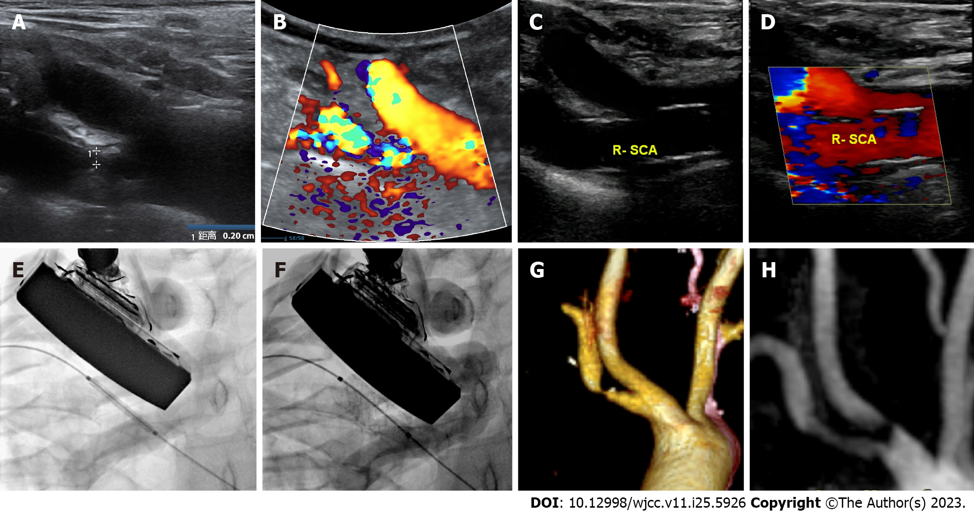
Figure 1 Images before and after subclavian artery stent implantation.
A: Ultrasound showing stenosis of the right subclavian artery (R-SCA) before surgery; B: Ultrasound showing blood flow in the region of R-SCA stenosis before surgery; C: Ultrasound showing good stent shape of the R-SCA after surgery; D: Ultrasound showing good blood flow in the region of R-SCA stent after surgery; E: X-ray showing the shape of the stent before stenting; F: X-ray showing the shape of the stent after stenting; G: Computed tomography angiography showing severe stenosis of the R-SCA before surgery; H: Magnetic resonance angiography showing good stent shape after surgery. R-SCA: Right subclavian artery.
- Citation: Li L, Wang ZY, Liu B. Ultrasound-guided carotid angioplasty and stenting in a patient with iodinated contrast allergy: A case report. World J Clin Cases 2023; 11(25): 5926-5933
- URL: https://www.wjgnet.com/2307-8960/full/v11/i25/5926.htm
- DOI: https://dx.doi.org/10.12998/wjcc.v11.i25.5926


